Questions and Answers ​in MRI
  • Home
  • Complete List of Questions
  • …Magnets & Scanners
    • Basic Electromagnetism >
      • What causes magnetism?
      • What is a Tesla?
      • Who was Tesla?
      • What is a Gauss?
      • How strong is 3.0T?
      • What is a gradient?
      • Aren't gradients coils?
      • What is susceptibility?
      • How to levitate a frog?
      • What is ferromagnetism?
      • Superparamagnetism?
    • Magnets - Part I >
      • Types of magnets?
      • Brands of scanners?
      • Which way does field point?
      • Which is the north pole?
      • Low v mid v high field?
      • Advantages to low-field?
      • Disadvantages?
      • What is homogeneity?
      • Why homogeneity?
      • Why shimming?
      • Passive shimming?
      • Active shimming?
    • Magnets - Part II >
      • Superconductivity?
      • Perpetual motion?
      • How to ramp?
      • Superconductive design?
      • Room Temp supercon?
      • Liquid helium use?
      • What is a quench?
      • Is field ever turned off?
      • Emergency stop button?
    • Gradients >
      • Gradient coils?
      • How do z-gradients work?
      • X- and Y- gradients?
      • Open scanner gradients?
      • Eddy current problems?
      • Active shielded gradients?
      • Active shield confusion?
      • What is pre-emphasis?
      • Gradient heating?
      • Gradient specifications?
      • Gradient linearity?
    • RF & Coils >
      • Many kinds of coils?
      • Radiofrequency waves?
      • Phase v frequency?
      • RF Coil function(s)?
      • RF-transmit coils?
      • LP vs CP (Quadrature)?
      • Multi-transmit RF?
      • Receive-only coils?
      • Array coils?
      • AIR Coils?
    • Site Planning >
      • MR system layout?
      • What are fringe fields?
      • How to reduce fringe?
      • Magnetic shielding?
      • Need for vibration testing?
      • What's that noise?
      • Why RF Shielding?
      • Wires/tubes thru wall?
  • ...Safety and Screening
    • Overview >
      • ACR Safety Zones?
      • MR safety screening?
      • Incomplete screening?
      • Passive v active implants?
      • Conditional implants?
      • Common safety issues?
      • Projectiles?
      • Metal detectors?
      • Pregnant patients?
      • Postop, ER & ICU patients?
      • Temperature monitoring?
      • Orbital foreign bodies?
      • Bullets and shrapnel?
    • Static Fields >
      • "Dangerous" metals?
      • "Safe" metals?
      • Magnetizing metal?
      • Object shape?
      • Forces on metal?
      • Most dangerous place?
      • Force/torque testing?
      • Static field bioeffects?
      • Dizziness/Vertigo?
      • Flickering lights?
      • Metallic taste?
    • RF Fields >
      • RF safety overview?
      • RF biological effects?
      • What is SAR?
      • SAR limits?
      • Operating modes?
      • How to reduce SAR?
      • RF burns?
      • Estimate implant heating?
      • SED vs SAR?
      • B1+rms vs SAR?
      • Personnel exposure?
      • Cell phones?
    • Gradient Fields >
      • Gradient safety overview
      • Acoustic noise?
      • Nerve stimulation?
      • Gradient vs RF heating?
    • Safety: Neurological >
      • Aneurysm coils/clips?
      • Shunts/drains?
      • Pressure monitors/bolts?
      • Deep brain stimulators?
      • Spinal cord stimulators?
      • Vagal nerve stimulators?
      • Cranial electrodes?
      • Carotid clamps?
      • Peripheral stimulators?
      • Epidural catheters?
    • Safety: Head & Neck >
      • Additional orbit safety?
      • Cochlear Implants?
      • Bone conduction implants?
      • Other ear implants?
      • Dental/facial implants?
      • ET tubes & airways?
    • Safety: Chest & Vascular >
      • Breast tissue expanders?
      • Breast biopsy markers?
      • Airway stents/valves/coils?
      • Respiratory stimulators?
      • Ports/vascular access?
      • Swan-Ganz catheters?
      • IVC filters?
      • Implanted infusion pumps?
      • Insulin pumps & CGMs?
      • Vascular stents/grafts?
      • Sternal wires/implants?
    • Safety: Cardiac >
      • Pacemaker dangers?
      • Pacemaker terminology?
      • New/'Safe" Pacemakers?
      • Old/Legacy Pacemakers?
      • Violating the conditions?
      • Epicardial pacers/leads?
      • Cardiac monitors?
      • Heart valves?
      • Miscellaneous CV devices?
    • Safety: Abdominal >
      • PIllCam and capsules?
      • Gastric pacemakers?
      • Other GI devices?
      • Contraceptive devices?
      • Foley catheters?
      • Incontinence devices?
      • Penile Implants?
      • Sacral nerve stimulators?
      • GU stents and other?
    • Safety: Orthopedic >
      • Orthopedic hardware?
      • External fixators?
      • Traction and halos?
      • Bone stimulators?
      • Magnetic rods?
  • …The NMR Phenomenon
    • Spin >
      • What is spin?
      • Why I = ½, 1, etc?
      • Proton = nucleus = spin?
      • Predict nuclear spin (I)?
      • Magnetic dipole moment?
      • Gyromagnetic ratio (γ)?
      • "Spin" vs "Spin state"?
      • Energy splitting?
      • Fall to lowest state?
      • Quantum "reality"?
    • Precession >
      • Why precession?
      • Who was Larmor?
      • Energy for precession?
      • Chemical shift?
      • Net magnetization (M)?
      • Does M instantly appear?
      • Does M also precess?
      • Does precession = NMR?
    • Resonance >
      • MR vs MRI vs NMR?
      • Who discovered NMR?
      • How does B1 tip M?
      • Why at Larmor frequency?
      • What is flip angle?
      • Spins precess after 180°?
      • Phase coherence?
      • Release of RF energy?
      • Rotating frame?
      • Off-resonance?
      • Adiabatic excitation?
      • Adiabatic pulses?
    • Relaxation - Physics >
      • Bloch equations?
      • What is T1?
      • What is T2?
      • Relaxation rate vs time?
      • Why is T1 > T2?
      • T2 vs T2*?
      • Causes of Relaxation?
      • Dipole-dipole interactions?
      • Chemical Exchange?
      • Spin-Spin interactions?
      • Macromolecule effects?
      • Which H's produce signal?
      • "Invisible" protons?
      • Magnetization Transfer?
      • Bo effect on T1 & T2?
      • How to predict T1 & T2?
    • Relaxation - Clincial >
      • T1 bright? - fat
      • T1 bright? - other oils
      • T1 bright? - cholesterol
      • T1 bright? - calcifications
      • T1 bright? - meconium
      • T1 bright? - melanin
      • T1 bright? - protein/mucin
      • T1 bright? - myelin
      • Magic angle?
      • MT Imaging/Contrast?
  • …Pulse Sequences
    • MR Signals >
      • Origin of MR signal?
      • Free Induction Decay?
      • Gradient echo?
      • TR and TE?
      • Spin echo?
      • 90°-90° Hahn Echo?
      • Stimulated echoes?
      • STEs for imaging?
      • 4 or more RF-pulses?
      • Partial flip angles?
      • How is signal higher?
      • Optimal flip angle?
    • Spin Echo >
      • SE vs Multi-SE vs FSE?
      • Image contrast: TR/TE?
      • Opposite effects ↑T1 ↑T2?
      • Meaning of weighting?
      • Does SE correct for T2?
      • Effect of 180° on Mz?
      • Direction of 180° pulse?
    • Inversion Recovery >
      • What is IR?
      • Why use IR?
      • Phase-sensitive IR?
      • Why not PSIR always?
      • Choice of IR parameters?
      • TI to null a tissue?
      • STIR?
      • T1-FLAIR
      • T2-FLAIR?
      • IR-prepped sequences?
      • Double IR?
    • Gradient Echo >
      • GRE vs SE?
      • Multi-echo GRE?
      • Types of GRE sequences?
      • Commercial Acronyms?
      • Spoiling - what and how?
      • Spoiled-GRE parameters?
      • Spoiled for T1W only?
      • What is SSFP?
      • GRASS/FISP: how?
      • GRASS/FISP: parameters?
      • GRASS vs MPGR?
      • PSIF vs FISP?
      • True FISP/FIESTA?
      • FIESTA v FIESTA-C?
      • DESS?
      • MERGE/MEDIC?
      • GRASE?
      • MP-RAGE v MR2RAGE?
    • Susceptibility Imaging >
      • What is susceptibility (χ)?
      • What's wrong with GRE?
      • Making an SW image?
      • Phase of blood v Ca++?
      • Quantitative susceptibility?
    • Diffusion: Basic >
      • What is diffusion?
      • Iso-/Anisotropic diffusion?
      • "Apparent" diffusion?
      • Making a DW image?
      • What is the b-value?
      • b0 vs b50?
      • Trace vs ADC map?
      • Light/dark reversal?
      • T2 "shine through"?
      • Exponential ADC?
      • T2 "black-out"?
      • DWI bright causes?
    • Diffusion: Advanced >
      • Diffusion Tensor?
      • DTI (tensor imaging)?
      • Whole body DWI?
      • Readout-segmented DWI?
      • Small FOV DWI?
      • IVIM?
      • Diffusion Kurtosis?
    • Fat-Water Imaging >
      • Fat & Water properties?
      • F-W chemical shift?
      • In-phase/out-of-phase?
      • Best method?
      • Dixon method?
      • "Fat-sat" pulses?
      • Water excitation?
      • STIR?
      • SPIR?
      • SPAIR v SPIR?
      • SPIR/SPAIR v STIR?
  • …Making an Image
    • From Signals to Images >
      • Phase v frequency?
      • Angular frequency (ω)?
      • Signal squiggles?
      • Real v Imaginary?
      • Fourier Transform (FT)?
      • What are 2D- & 3D-FTs?
      • Who invented MRI?
      • How to locate signals?
    • Frequency Encoding >
      • Frequency encoding?
      • Receiver bandwidth?
      • Narrow bandwidth?
      • Slice-selective excitation?
      • SS gradient lobes?
      • Cross-talk?
      • Frequency encode all?
      • Mixing of slices?
      • Two slices at once?
      • Simultaneous Multi-Slice?
    • Phase Encoding >
      • Phase-encoding gradient?
      • Single PE step?
      • What is phase-encoding?
      • PE and FE together?
      • 2DFT reconstruction?
      • Choosing PE/FE direction?
    • Performing an MR Scan >
      • What are the steps?
      • Automatic prescan?
      • Routine shimming?
      • Coil tuning/matching?
      • Center frequency?
      • Transmitter gain?
      • Receiver gain?
      • Dummy cycles?
      • Where's my data?
      • MR Tech qualifications?
    • Image Quality Control >
      • Who regulates MRI?
      • Who accredits?
      • Mandatory accreditation?
      • Routine quality control?
      • MR phantoms?
      • Geometric accuracy?
      • Image uniformity?
      • Slice parameters?
      • Image resolution?
      • Signal-to-noise?
      • Ghosting?
  • …K-space & Rapid Imaging
    • K-space (Basic) >
      • What is k-space?
      • Parts of k-space?
      • What does "k" stand for?
      • Spatial frequencies?
      • Locations in k-space?
      • Data for k-space?
      • Why signal ↔ k-space?
      • Spin-warp imaging?
      • Big spot in middle?
      • K-space trajectories?
      • Radial sampling?
    • K-space (Advanced) >
      • K-space grid?
      • Negative frequencies?
      • Field-of-view (FOV)
      • Rectangular FOV?
      • Partial Fourier?
      • Phase symmetry?
      • Read symmetry?
      • Why not use both?
      • ZIP?
    • Rapid Imaging (FSE &EPI) >
      • What is FSE/TSE?
      • FSE parameters?
      • Bright Fat?
      • Other FSE differences?
      • Dual-echo FSE?
      • Driven equilibrium?
      • Reduced flip angle FSE?
      • Hyperechoes?
      • SPACE/CUBE/VISTA?
      • Echo-planar imaging?
      • HASTE/SS-FSE?
    • Parallel Imaging (PI) >
      • What is PI?
      • How is PI different?
      • PI coils and sequences?
      • Why and when to use?
      • Two types of PI?
      • SENSE/ASSET?
      • GRAPPA/ARC?
      • CAIPIRINHA?
      • Compressed sensing?
      • Noise in PI?
      • Artifacts in PI?
  • …Contrast Agents
    • Contrast Agents: Physics >
      • Why Gadolinium?
      • Paramagnetic relaxation?
      • What is relaxivity?
      • Why does Gd shorten T1?
      • Does Gd affect T2?
      • Gd & field strength?
      • Best T1-pulse sequence?
      • Triple dose and MT?
      • Dynamic CE imaging?
      • Gadolinium on CT?
    • Contrast Agents: Clinical >
      • So many Gd agents!
      • Important properties?
      • Ionic v non-ionic?
      • Intra-articular/thecal Gd?
      • Gd liver agents (Eovist)?
      • Mn agents (Teslascan)?
      • Feridex & Liver Agents?
      • Lymph node agents?
      • Ferumoxytol?
      • Blood pool (Ablavar)?
      • Bowel contrast agents?
    • Contrast Agents: Safety >
      • Gadolinium safety?
      • Allergic reactions?
      • Renal toxicity?
      • What is NSF?
      • NSF by agent?
      • Informed consent for Gd?
      • Gd protocol?
      • Is Gd safe in infants?
      • Reduced dose in infants?
      • Gd in breast milk?
      • Gd in pregnancy?
      • Gd accumulation?
      • Gd deposition disease?
  • …Cardiovascular and MRA
    • Flow effects in MRI >
      • Defining flow?
      • Expected velocities?
      • Laminar v turbulent?
      • Predicting MR of flow?
      • Time-of-flight effects?
      • Spin phase effects?
      • Flow void?
      • Why GRE ↑ flow signal?
      • Slow flow v thrombus?
      • Even-echo rephasing?
      • Flow-compensation?
      • Flow misregistration?
    • MR Angiography - I >
      • MRA methods?
      • Dark vs bright blood?
      • Time-of-Flight (TOF) MRA?
      • 2D vs 3D MRA?
      • MRA parameters?
      • Magnetization Transfer?
      • Ramped flip angle?
      • MOTSA?
      • Fat-suppressed MRA?
      • TOF MRA Artifacts?
      • Phase-contrast MRA?
      • What is VENC?
      • Measuring flow?
      • 4D Flow Imaging?
      • How accurate?
    • MR Angiography - II >
      • Gated 3D FSE MRA?
      • 3D FSE MRA parameters?
      • SSFP MRA?
      • Inflow-enhanced SSFP?
      • MRA with ASL?
      • Other MRA methods?
      • Contrast-enhanced MRA?
      • Timing the bolus?
      • View ordering in MRA?
      • Bolus chasing?
      • TRICKS or TWIST?
      • CE-MRA artifacts?
    • Cardiac I - Intro/Anatomy >
      • Cardiac protocols?
      • Patient prep?
      • EKG problems?
      • Magnet changes EKG?
      • Gating v triggering?
      • Gating parameters?
      • Heart navigators?
      • Dark blood/Double IR?
      • Why not single IR?
      • Triple IR?
      • Polar plots?
      • Coronary artery MRA?
    • Cardiac II - Function >
      • Beating heart movies?
      • Cine parameters?
      • Real-time cine?
      • Ventricular function?
      • Tagging/SPAMM?
      • Perfusion: why and how?
      • 1st pass perfusion?
      • Quantifying perfusion?
      • Dark rim artifact
    • Cardiac III - Viability >
      • Gd enhancement?
      • TI to null myocardium?
      • PS (phase-sensitive) IR?
      • Wideband LGE?
      • T1 mapping?
      • Iron/T2*-mapping?
      • Edema/T2-mapping?
      • Why/how stress test?
      • Stess drugs/agents?
      • Stress consent form?
  • …MR Artifacts
    • Tissue-related artifacts >
      • Chemical shift artifact?
      • Chemical shift in phase?
      • Reducing chemical shift?
      • Chemical Shift 2nd Kind?
      • In-phase/out-of phase?
      • IR bounce point?
      • Susceptibility artifact?
      • Metal suppression?
      • Dielectric effect?
      • Dielectric Pads?
    • Motion-related artifacts >
      • Why discrete ghosts?
      • Motion artifact direction?
      • Reducing motion artifacts?
      • Saturation pulses?
      • Gating methods?
      • Respiratory comp?
      • Navigator echoes?
      • PROPELLER/BLADE?
    • Technique-related artifacts >
      • Partial volume effects?
      • Slice overlap?
      • Aliasing?
      • Wrap-around artifact?
      • Eliminate wrap-around?
      • Phase oversampling?
      • Frequency wrap-around?
      • Spiral/radial artifacts?
      • Gibbs artifact?
      • Nyquist (N/2) ghosts?
      • Zipper artifact?
      • Data artifacts?
      • Surface coil flare?
      • MRA Artifacts (TOF)?
      • MRA artifacts (CE)?
  • …Functional Imaging
    • Perfusion I: Intro & DSC >
      • Measuring perfusion?
      • Meaning of CBF, MTT etc?
      • DSC v DCE v ASL?
      • How to perform DSC?
      • Bolus Gd effect?
      • T1 effects on DSC?
      • DSC recirculation?
      • DSC curve analysis?
      • DSC signal v [Gd]
      • Arterial input (AIF)?
      • Quantitative DSC?
    • Perfusion II: DCE >
      • What is DCE?
      • How is DCE performed?
      • How is DCE analyzed?
      • Breast DCE?
      • DCE signal v [Gd]
      • DCE tissue parmeters?
      • Parameters to images?
      • K-trans = permeability?
      • Utility of DCE?
    • Perfusion III: ASL >
      • What is ASL?
      • ASL methods overview?
      • CASL?
      • PASL?
      • pCASL?
      • ASL parameters?
      • ASL artifacts?
      • Gadolinium and ASL?
      • Vascular color maps?
      • Quantifying flow?
    • Functional MRI/BOLD - I >
      • Who invented fMRI?
      • How does fMRI work?
      • BOLD contrast?
      • Why does BOLD ↑ signal?
      • Does BOLD=brain activity?
      • BOLD pulse sequences?
      • fMRI Paradigm design?
      • Why "on-off" comparison?
      • Motor paradigms?
      • Visual?
      • Language?
    • Functional MRI/BOLD - II >
      • Process/analyze fMRI?
      • Best fMRI software?
      • Data pre-processing?
      • Registration/normalization?
      • fMRI statistical analysis?
      • General Linear Model?
      • Activation "blobs"?
      • False activation?
      • Resting state fMRI?
      • Analyze RS-fMRI?
      • Network/Graphs?
      • fMRI at 7T?
      • Mind reading/Lie detector?
      • fMRI critique?
  • …MR Spectroscopy
    • MRS I - Basics >
      • MRI vs MRS?
      • Spectra vs images?
      • Chemical shift (δ)?
      • Measuring δ?
      • Backward δ scale?
      • Predicting δ?
      • Size/shapes of peaks?
      • Splitting of peaks?
      • Localization methods?
      • Single v multi-voxel?
      • PRESS?
      • STEAM?
      • ISIS?
      • CSI?
    • MRS II - Clinical ¹H MRS >
      • How-to: brain MRS?
      • Water suppression?
      • Fat suppression?
      • Normal brain spectra?
      • Choice of TR/TE/etc?
      • Hunter's angle?
      • Lactate inversion?
      • Metabolite mapping?
      • Metabolite quantitation?
      • Breast MRS?
      • Gd effect on MRS?
      • How-to: prostate MRS?
      • Prostate spectra?
      • Muscle ¹H-MRS?
      • Liver ¹H-MRS?
      • MRS artifacts?
    • MRS III - Multi-nuclear >
      • Other nuclei?
      • Why phosphorus?
      • How-to: ³¹P MRS
      • Normal ³¹P spectra?
      • Organ differences?
      • ³¹P measurements?
      • Decoupling?
      • NOE?
      • Carbon MRS?
      • Sodium imaging?
      • Xenon imaging?
  • ...Artificial Intelligence
    • AI Part I: Basics >
      • Artificial Intelligence (AI)?
      • What is a neural network?
      • Machine Learning (ML)?
      • Shallow v Deep ML?
      • Shallow networks?
      • Deep network types?
      • Data prep and fitting?
      • Back-Propagation?
      • DL 'Playground'?
    • AI Part 2: Advanced >
      • What is convolution?
      • Convolutional Network?
      • Softmax?
      • Upsampling?
      • Limitations/Problems of AI?
      • Is the Singularity near?
    • AI Part 3: Image processing >
      • AI in clinical MRI?
      • Super-resolution?
  • ...Tissue Properties Imaging
    • MRI of Hemorrhage >
      • Hematoma overview?
      • Types of Hemoglobin?
      • Hyperacute/Oxy-Hb?
      • Acute/Deoxy-Hb?
      • Subacute/Met-Hb?
      • Deoxy-Hb v Met-Hb?
      • Extracellular met-Hb?
      • Chronic hematomas?
      • Hemichromes?
      • Ferritin/Hemosiderin?
      • Subarachnoid blood?
      • Blood at lower fields?
    • T2 cartilage mapping
    • MR Elastography?
    • Synthetic MRI?
    • Amide Proton Transfer?
    • MR thermography?
    • Electric Properties Imaging?
  • Copyright/Legal
    • Copyright Issues
    • Legal Disclaimers
  • Forums/Blogs/Links
  • What's New
  • Self-test Quizzes - NEW!
    • Magnets & Scanners Quiz
    • Safety & Screening Quiz
    • NMR Phenomenon Quiz
    • Pulse Sequences Quiz
    • Making an Image Quiz
    • K-space & Rapid Quiz
    • Contrast & Blood Quiz
    • Cardiovascular & MRA Quiz

Additional Orbital Safety Issues

Besides foreign bodies, are there any other eye-related potential hazards to be aware of in MRI? 
orbit safety mri
Read MR Safety Disclaimer Before Proceeding
Yes. Several immediately come to mind.
Cosmetics
All eye make-up, especially mascara, should be removed prior to imaging.  Such cosmetics often contain iron oxides that create a local artifact and may even result in eye irritation during MR imaging.
Permanent eye-liner and periorbital tattoos may cause local swelling and pain even hours after scanning. Although some experts have recommended placing cold packs on the eyes preemptively, we simply warn the patient about this possibility and tell them to notify us immediately if any discomfort is experienced.
mascara artifact MRI
Susceptibility artifact from iron particles in mascara
Recently magnetic artificial eyelashes have become available which constitute a potential MR safety hazard as well as a source of artifacts. Instead of glue, the user paints on a black eyeliner containing ultrafine magnetic particles. The lashes themselves contain thin magnetic strips at their bases to attach to the lids. Both the lashes and eyeliner should be removed prior to imaging.
Artificial Lenses
Virtually all intraocular lens (IOL) implants are made entirely of plastic or silicone, and thus present no safety concerns in the MR environment. In a few older models, metallic loops made of platinium, iridium or titanium may be attached to the lens for suturing purposes; however, all tested to date have demonstrated no significant heating or displacement at field strengths up to 7.0T. One relatively new complex IOL, the implantable miniature telescope, is considered MR Conditional because it contains a small stainless steel part in its center, so there are restrictions on maximum field and gradient strength.
lens implant
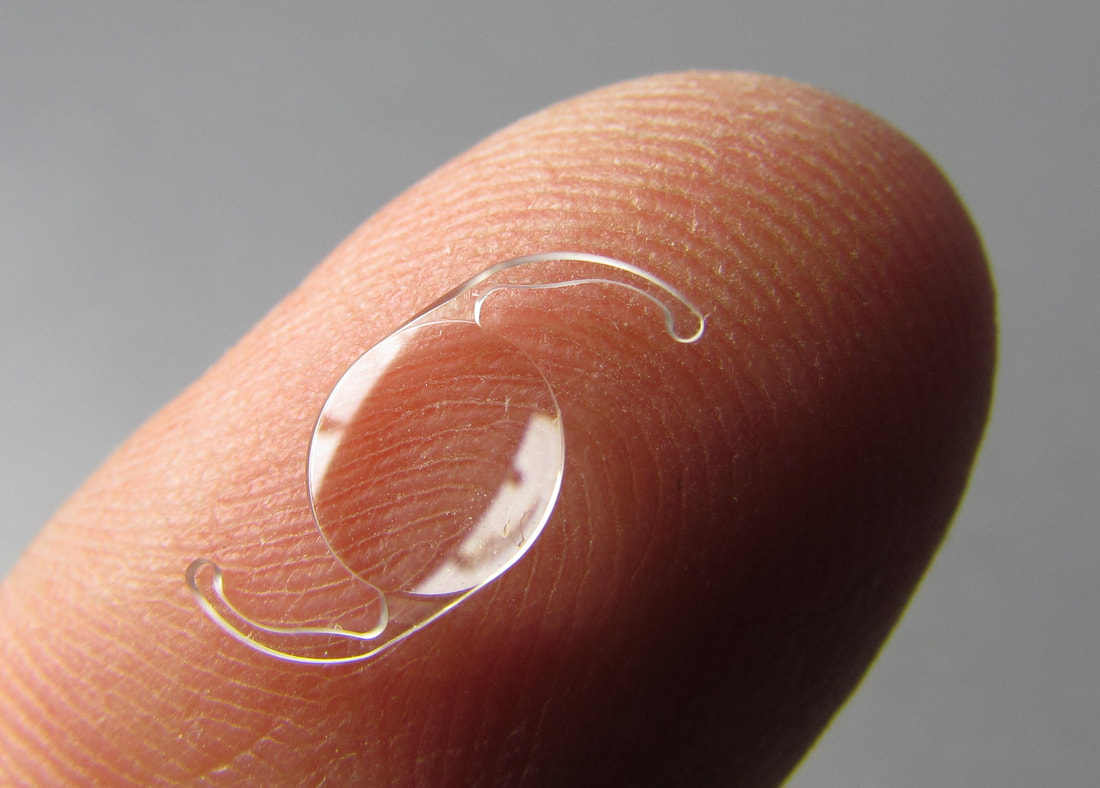
Acrylic Intraocular Lens Implant
Worst lens implant
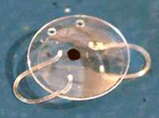
Worst Medallion Implant with metal loops
implantable telescope lens
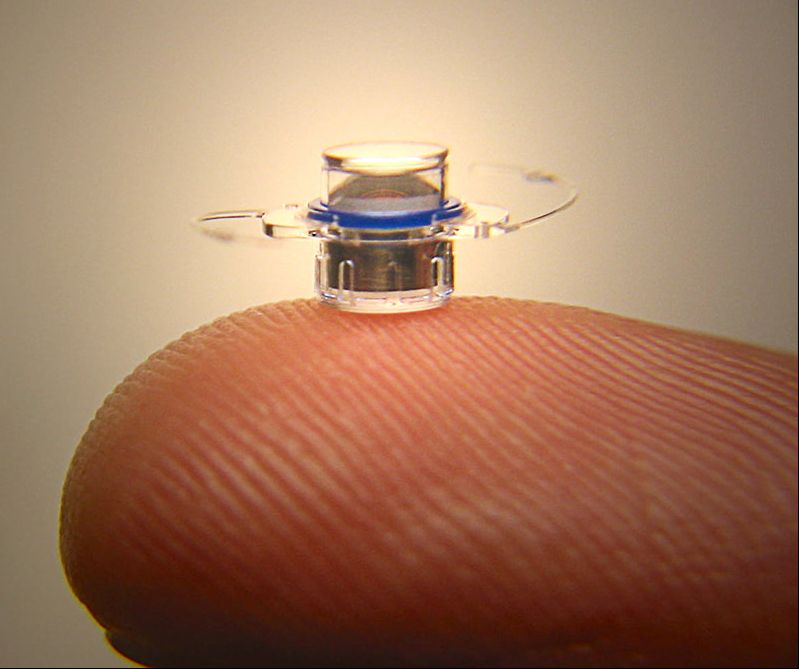
VisionCare implantable miniature telescope
Conventional external contact lenses are also entirely safe, even the new fully implantable ones. It should be noted that certain cosmetic "circle" lenses contain iron oxide pigments that can produce a localized artifact on MR images (but without apparent adverse health effects).
The single MR Unsafe implant in this category is the relatively new Sensimed Triggerfish, a contact lens with an embedded micro-sensor that captures spontaneous circumferential changes at the corneoscleral area (an indirect index for anterior chamber size and hence intraocular pressure).  These uncommonly used devices are implanted for a 24-hour period to continuously monitor intraocular pressure in glaucoma patients, then removed.
Picture
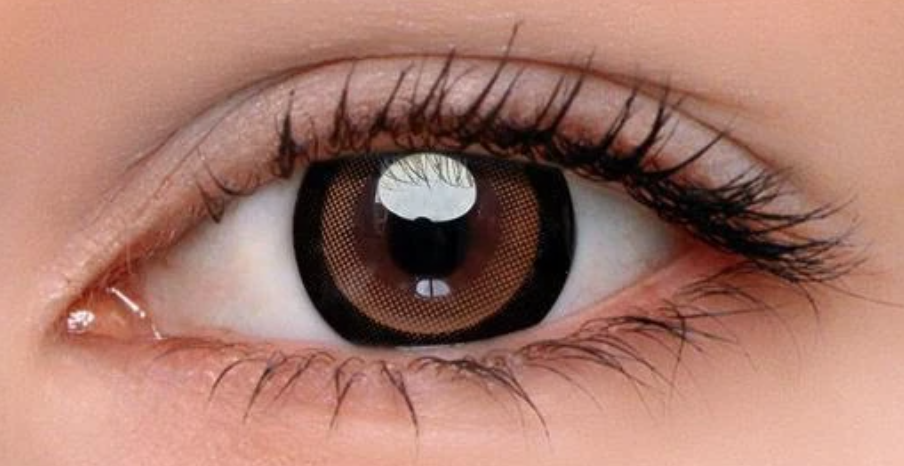
Black "circle" contact lens
Triggerfish Sensimed
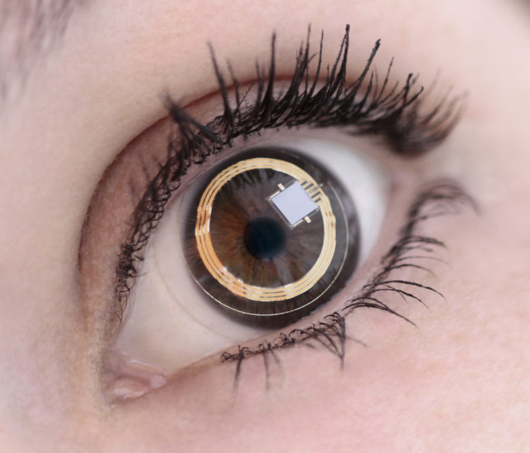
MR Unsafe Triggerfish contact lens
Eyelid Weights and Springs
Internal palpebral weights made of gold, platinum, or platinum-iridium and are inserted in the pretarsal space of the upper lid to passively assist with lid closure in patients with facial nerve palsy. Although most are probably safe even up to 7.0T, some are labeled MR Conditional by manufacturers (out of an abundance of caution, I suppose). Externally applied weights are also available, such as the Blinkeze™ plate held in place by an adhesive strip. Even though made of (non-ferromagnetic) tantalum, such external weights should be removed prior to MRI to eliminate the possibility of skin burns.
Picture
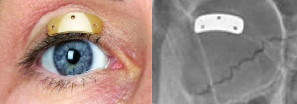
Gold weight positioned over upper lid prior to surgery and postoperative orbit x-ray
Picture
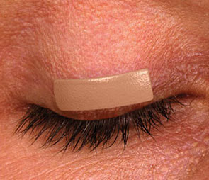
Blinkeze™ external eyelid weight
Implanted palpebral springs, more widely used in the 1970s and 1980s than today, assist with active closure of the lid. These devices are not commercially produced, but each is individually crafted by the surgeon using stainless steel, Nitinol (nickel-titanium), or nonferrous 35 NLT alloy wire. Although some concerns about movement within 1 month of placement and localized discomfort have been voiced, many patients have undergone uneventful MRI at 3T or less with the only precaution being placement of a protective plastic covering and eye patch. 
Morel-Fatio eyelid spring
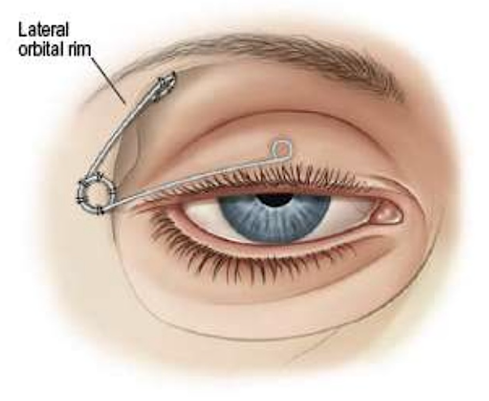
Morel-Fatio palpebral spring
Glaucoma Devices
EX-PRESS glaucoma deviceMetal EX-PRESS glaucoma shunt
Implanted glaucoma devices are used to reduce intraocular pressure by removing aqueous humor from the anterior chamber of the eye. They are virtually all constructed out of silicone or plastic and are considered MR Safe (with two exceptions):  

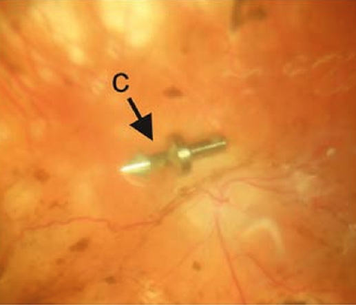
​The first exception is the metal EX-PRESS Glaucoma Filtration Device, a 2-3 mm long pointed stainless steel tube that is implanted under a scleral flap. The tube drains fluid from the anterior chamber into the intrascleral space. The EX-PRESS mimics a foreign body on x-ray and creates a local artifact on MR. It is considered MR Conditional up to 3.0T with the additional caveat that the manufacturer does not recommend scanning in the first 2 weeks post implantation.

Hydrus® microstent
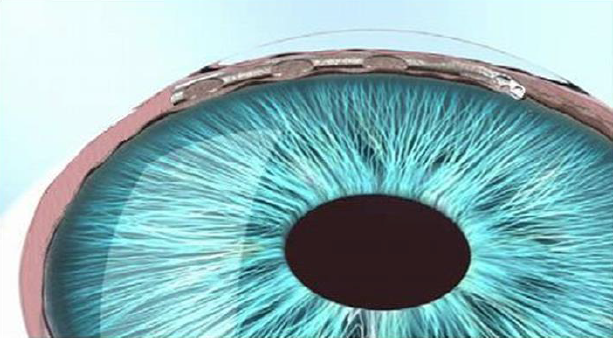
"MR conditional" Hydrus® microstent
The second exception is the Hydrus® Microstent (Ivantis), a crescent-shaped device made of non-ferromagnetic nitinol (an alloy of nickel and titanium) placed in Schlemm’s canal near the corneal-iris junction. (Schlemm’s canal is a lymphatic-like vessel in the deep cornea whose function is to drain aqueous humor from the anterior chamber into the episcleral blood vessels). Like the EX-PRESS Device, the Hydrus is MR Conditional up to 3.0T.

Scleral Banding
Scleral buckling or banding is used to treat chronic retinal detachment. The procedure involves encircling the globe with a belt, band, buckle, or tire-like structure made of silicone or hydrogel, occasionally held in place by metallic clip. For the last 30 years, these clips have been made of tantalum, and hence pose no risk of movement in an external magnetic field.
Picture
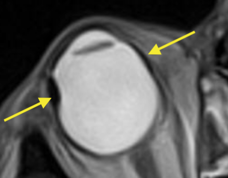
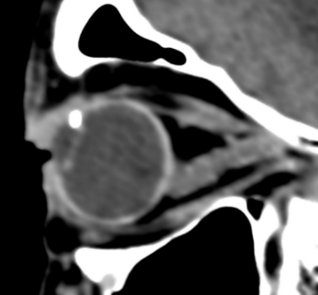
T2-weighted image showing scleral band
Eye Prostheses
hydroxyapatite eye prosthesis
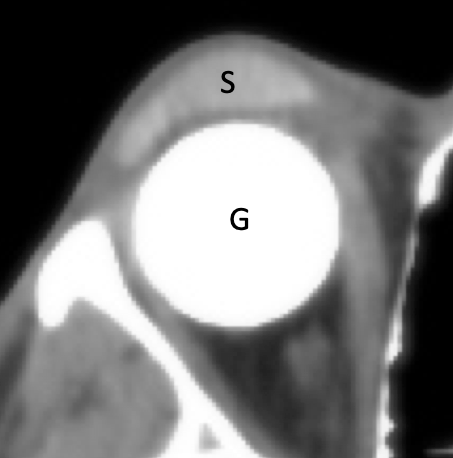
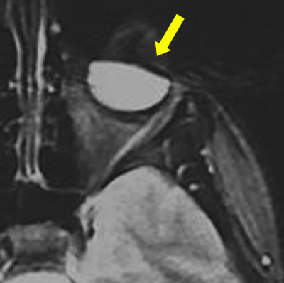
Eye prosthesis with globe (G) and scleral shell (S). The globe is dense due to its hydroxyapatite composition, not to be construed as metallic.
The typical prosthetic eye consists of: 1) a posterior globe component, commonly made of hydroxyapatite, porous polyethlyene, acrylic, silicone, or bioceramic; and 2) a removable anterior shell, made of acrylic or occasionally glass and painted to resemble the normal eye. The extraocular muscles may or may not be sutured to the implant. Some patients have only a scleral shell without a posterior globe prosthesis. Because none of these components in standard eye prostheses contain metal, they are all considered MR Safe. Some eye prostheses have a titanium peg that couples the anterior and posterior components so they move together. These are also "MR safe". 
As a warning, a small number of legacy MEDPOR prostheses implanted in the 2000-2010 time frame that contain a small magnet in the shell magnetically coupled to a ferromagnetic screw on the globe component. MR scanning of these prostheses is contraindicated. 
Miscellaneous and Uncommon Devices
Picture
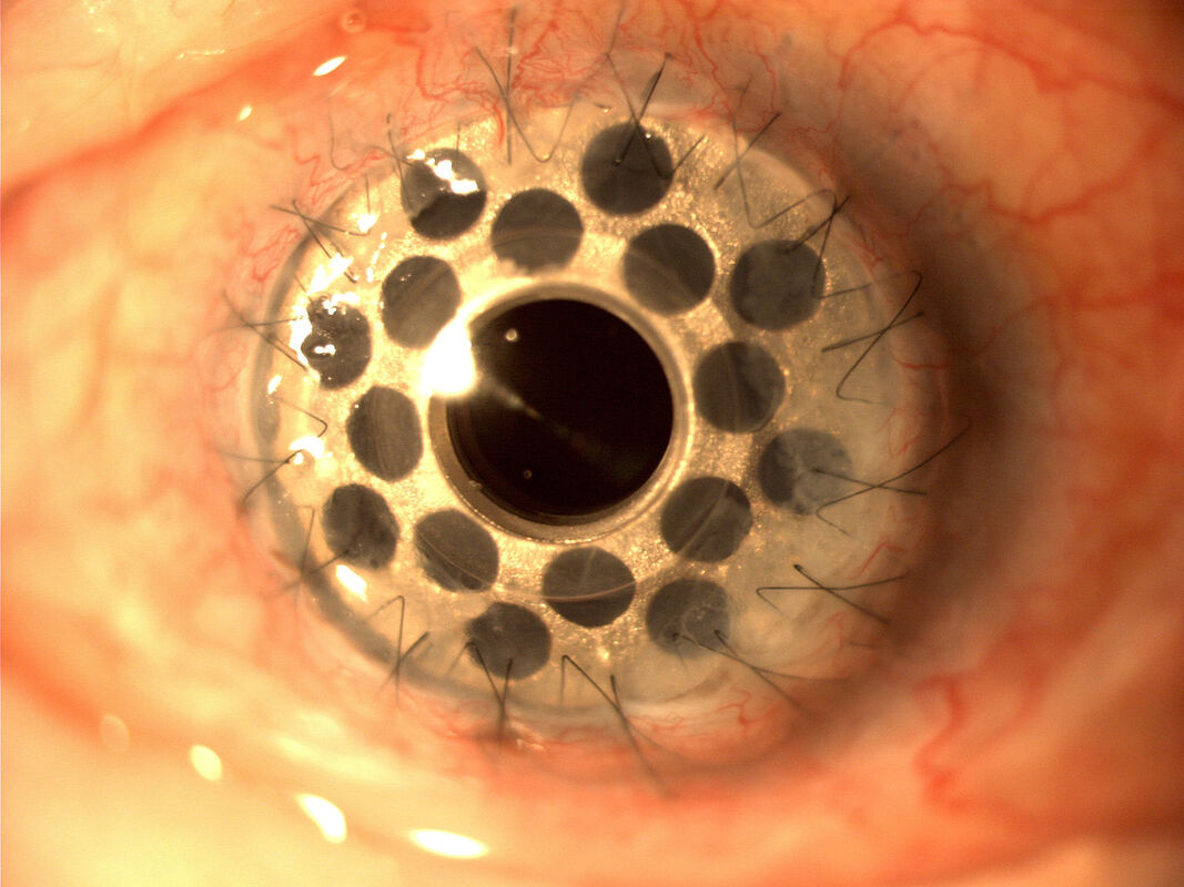
Boston Keratoprosthesis (MRI Conditional)
Corneal Implants. The Boston Kerato-prosthesis is designed as an artificial cornea for patients with previous failed graft attempts. The collar-button-like device attaches to the native cornea and contains consists of a clear sheet of plastic holding a donor graft and affixed to a back plate with locking titanium ring. Because of the possible tissue heating near the metal component, the device is considered MR Conditional.
​'Electronic Retinal Implants. Two devices, the Argus II Retinal Prosthesis System and the IRIS II have been granted CE mark for use in Europe (the Argus also has FDA approval in the US). Both devices comprise an epiretinal electrode array located on the surface of the retina together with an eyeglass-mounted digital camera transmitting signals wirelessly to the impant's receiver. With the external hardware removed, both devices are considered MR Conditional at fields up to 3.0T.
Argus II Retinal Prosthesis
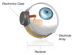
Argus II Retinal Prosthesis (MRI Conditional)
Picture
Extruded retinal tack (from Schwering under CC BY)
Retinal tacks made of plastic or metal were reasonably popular in the 1980s as an adjunct for repair of complicated retinal detachments. At least one of these tacks, the Western European model, was made of ferromagnetic steel and has long been singled out as MR Unsafe. Retinal tacks are seldom used today because of their general commercial unavailability and associated complications. However, the Argus II Retinal Prosthesis uses a single titanium retinal tack to hold the electrode array in place and is MR Conditional.

Advanced Discussion (show/hide)»

No supplementary material yet. Check back soon!

References     
     Albert DW, Olsen KR Parel J-M, et al. Magnetic resonance imaging and retinal tacks (letter). Arch Ophthalmol 1990; 108:320-321.
     Bakshandeh H, Shellock FG, Schatz CJ, Morisoli SM. Metallic clips used for scleral buckling: ex vivo evaluation of ferromagnetism at 1.5T (letter). J Magn Reson Imaging 1993: 3:559. (they are safe)
     Luo YH-L, da Cruz L. A review and update on the current status of retinal prostheses (bionic eye). Br Med Bull 2014; 109:31-44. [DOI Link]
     Mabray MC, Uzelac A, Talbott JF, et al. Ex-PRESS glaucoma filter: an MRI compatible metallic orbital foreign body imaged at 1.5 and 3 T. Clin Radiol 2015; 70:e28-e34.  [DOI LINK]
     Reiter MJ, Schwope RB, Kini JA, et al. Postoperative imaging of the orbital contents. RadioGraphics 2015; 35:221-234. [DOI LINK]
     Schrom T, Thelen A, Asbach P, Bauknecht H-C. Effect of 7.0 Tesla MRI on upper eyelid implants. Ophthalm Plast Recon Surg 2006; 22:480-482. [DOI Link]  (no heating or displacement for three types of eyelid weights tested at 7.0T)
     
Schwering MS, Oltrup T, Rückheim KS, et al. New retinal tack designs: an analysis of retention forces in human scleral tissue. Graefe's Arch Clin Exp Ophthalmol 2020; 258:1389-1394. [DOI Link]
     Seiff SR, Vestel KP, Truwit CL. Eyelid palpebral springs in patients undergoing magnetic resonance imaging: an area of possible concern (letter). Arch Ophthalmol 1991; 109:319.
     
Slonimsky E, Mamourian A. Magnetic eyelashes: a new source of MRI artifacts. AJR Am J Roentgenol 2019; 213:983-985.
     van Rijn GA, Mourik JEM, Teeuwisse WM, et al. Magnetic resonance compatibility of intraocular lenses measured at 7 Tesla. IOVS Invest Ophthalmol Visual Sci 2012; 53:3449-53. [DOI LINK] 
​     
Weiland JD, Faraji B, Greenberg Rj, et al. Assessment of MRI issues for the Argus II Retinal Prosthesis. Magn Reson Imaging 2012; 30:382-389.
     Yuh WTC, Hanigan MT, Nerad JA, et al. Extrusion of eye socket magnetic implant after MR imaging: potential hazard to patient with eye prosthesis. J Magn Reson Imaging 1991; 1:711-2. (included for historical interest only; the Troutman extruded magnetic implant was used mostly between 1940 and 1960, so there are probably no patients remaining alive with this device).

Related Questions
     I've heard people have been blinded by metallic foreign bodies in the eye. How do you screen for these?
     How do you screen patients for implants and foreign bodies prior to MRI?

←  Previous Question
Next Question  →
↑ Complete List of Questions ↑
© 2024 AD Elster, ELSTER LLC
All rights reserved.   
MRIquestions.com - Home
Donate
Please help keep this site free for everyone in the world!