Questions and Answers ​in MRI
  • Home
  • Complete List of Questions
  • …Magnets & Scanners
    • Basic Electromagnetism >
      • What causes magnetism?
      • What is a Tesla?
      • Who was Tesla?
      • What is a Gauss?
      • How strong is 3.0T?
      • What is a gradient?
      • Aren't gradients coils?
      • What is susceptibility?
      • How to levitate a frog?
      • What is ferromagnetism?
      • Superparamagnetism?
    • Magnets - Part I >
      • Types of magnets?
      • Brands of scanners?
      • Which way does field point?
      • Which is the north pole?
      • Low v mid v high field?
      • Advantages to low-field?
      • Disadvantages?
      • What is homogeneity?
      • Why homogeneity?
      • Why shimming?
      • Passive shimming?
      • Active shimming?
    • Magnets - Part II >
      • Superconductivity?
      • Perpetual motion?
      • How to ramp?
      • Superconductive design?
      • Room Temp supercon?
      • Liquid helium use?
      • What is a quench?
      • Is field ever turned off?
      • Emergency stop button?
    • Gradients >
      • Gradient coils?
      • How do z-gradients work?
      • X- and Y- gradients?
      • Open scanner gradients?
      • Eddy current problems?
      • Active shielded gradients?
      • Active shield confusion?
      • What is pre-emphasis?
      • Gradient heating?
      • Gradient specifications?
      • Gradient linearity?
    • RF & Coils >
      • Many kinds of coils?
      • Radiofrequency waves?
      • Phase v frequency?
      • RF Coil function(s)?
      • RF-transmit coils?
      • LP vs CP (Quadrature)?
      • Multi-transmit RF?
      • Receive-only coils?
      • Array coils?
      • AIR Coils?
    • Site Planning >
      • MR system layout?
      • What are fringe fields?
      • How to reduce fringe?
      • Magnetic shielding?
      • Need for vibration testing?
      • What's that noise?
      • Why RF Shielding?
      • Wires/tubes thru wall?
  • ...Safety and Screening
    • Overview >
      • ACR Safety Zones?
      • MR safety screening?
      • Incomplete screening?
      • Passive v active implants?
      • Conditional implants?
      • Common safety issues?
      • Projectiles?
      • Metal detectors?
      • Pregnant patients?
      • Postop, ER & ICU patients?
      • Temperature monitoring?
      • Orbital foreign bodies?
      • Bullets and shrapnel?
    • Static Fields >
      • "Dangerous" metals?
      • "Safe" metals?
      • Magnetizing metal?
      • Object shape?
      • Forces on metal?
      • Most dangerous place?
      • Force/torque testing?
      • Static field bioeffects?
      • Dizziness/Vertigo?
      • Flickering lights?
      • Metallic taste?
    • RF Fields >
      • RF safety overview?
      • RF biological effects?
      • What is SAR?
      • SAR limits?
      • Operating modes?
      • How to reduce SAR?
      • RF burns?
      • Estimate implant heating?
      • SED vs SAR?
      • B1+rms vs SAR?
      • Personnel exposure?
      • Cell phones?
    • Gradient Fields >
      • Gradient safety overview
      • Acoustic noise?
      • Nerve stimulation?
      • Gradient vs RF heating?
    • Safety: Neurological >
      • Aneurysm coils/clips?
      • Shunts/drains?
      • Pressure monitors/bolts?
      • Deep brain stimulators?
      • Spinal cord stimulators?
      • Vagal nerve stimulators?
      • Cranial electrodes?
      • Carotid clamps?
      • Peripheral stimulators?
      • Epidural catheters?
    • Safety: Head & Neck >
      • Additional orbit safety?
      • Cochlear Implants?
      • Bone conduction implants?
      • Other ear implants?
      • Dental/facial implants?
      • ET tubes & airways?
    • Safety: Chest & Vascular >
      • Breast tissue expanders?
      • Breast biopsy markers?
      • Airway stents/valves/coils?
      • Respiratory stimulators?
      • Ports/vascular access?
      • Swan-Ganz catheters?
      • IVC filters?
      • Implanted infusion pumps?
      • Insulin pumps & CGMs?
      • Vascular stents/grafts?
      • Sternal wires/implants?
    • Safety: Cardiac >
      • Pacemaker dangers?
      • Pacemaker terminology?
      • New/'Safe" Pacemakers?
      • Old/Legacy Pacemakers?
      • Violating the conditions?
      • Epicardial pacers/leads?
      • Cardiac monitors?
      • Heart valves?
      • Miscellaneous CV devices?
    • Safety: Abdominal >
      • PIllCam and capsules?
      • Gastric pacemakers?
      • Other GI devices?
      • Contraceptive devices?
      • Foley catheters?
      • Incontinence devices?
      • Penile Implants?
      • Sacral nerve stimulators?
      • GU stents and other?
    • Safety: Orthopedic >
      • Orthopedic hardware?
      • External fixators?
      • Traction and halos?
      • Bone stimulators?
      • Magnetic rods?
  • …The NMR Phenomenon
    • Spin >
      • What is spin?
      • Why I = ½, 1, etc?
      • Proton = nucleus = spin?
      • Predict nuclear spin (I)?
      • Magnetic dipole moment?
      • Gyromagnetic ratio (γ)?
      • "Spin" vs "Spin state"?
      • Energy splitting?
      • Fall to lowest state?
      • Quantum "reality"?
    • Precession >
      • Why precession?
      • Who was Larmor?
      • Energy for precession?
      • Chemical shift?
      • Net magnetization (M)?
      • Does M instantly appear?
      • Does M also precess?
      • Does precession = NMR?
    • Resonance >
      • MR vs MRI vs NMR?
      • Who discovered NMR?
      • How does B1 tip M?
      • Why at Larmor frequency?
      • What is flip angle?
      • Spins precess after 180°?
      • Phase coherence?
      • Release of RF energy?
      • Rotating frame?
      • Off-resonance?
      • Adiabatic excitation?
      • Adiabatic pulses?
    • Relaxation - Physics >
      • Bloch equations?
      • What is T1?
      • What is T2?
      • Relaxation rate vs time?
      • Why is T1 > T2?
      • T2 vs T2*?
      • Causes of Relaxation?
      • Dipole-dipole interactions?
      • Chemical Exchange?
      • Spin-Spin interactions?
      • Macromolecule effects?
      • Which H's produce signal?
      • "Invisible" protons?
      • Magnetization Transfer?
      • Bo effect on T1 & T2?
      • How to predict T1 & T2?
    • Relaxation - Clincial >
      • T1 bright? - fat
      • T1 bright? - other oils
      • T1 bright? - cholesterol
      • T1 bright? - calcifications
      • T1 bright? - meconium
      • T1 bright? - melanin
      • T1 bright? - protein/mucin
      • T1 bright? - myelin
      • Magic angle?
      • MT Imaging/Contrast?
  • …Pulse Sequences
    • MR Signals >
      • Origin of MR signal?
      • Free Induction Decay?
      • Gradient echo?
      • TR and TE?
      • Spin echo?
      • 90°-90° Hahn Echo?
      • Stimulated echoes?
      • STEs for imaging?
      • 4 or more RF-pulses?
      • Partial flip angles?
      • How is signal higher?
      • Optimal flip angle?
    • Spin Echo >
      • SE vs Multi-SE vs FSE?
      • Image contrast: TR/TE?
      • Opposite effects ↑T1 ↑T2?
      • Meaning of weighting?
      • Does SE correct for T2?
      • Effect of 180° on Mz?
      • Direction of 180° pulse?
    • Inversion Recovery >
      • What is IR?
      • Why use IR?
      • Phase-sensitive IR?
      • Why not PSIR always?
      • Choice of IR parameters?
      • TI to null a tissue?
      • STIR?
      • T1-FLAIR
      • T2-FLAIR?
      • IR-prepped sequences?
      • Double IR?
    • Gradient Echo >
      • GRE vs SE?
      • Multi-echo GRE?
      • Types of GRE sequences?
      • Commercial Acronyms?
      • Spoiling - what and how?
      • Spoiled-GRE parameters?
      • Spoiled for T1W only?
      • What is SSFP?
      • GRASS/FISP: how?
      • GRASS/FISP: parameters?
      • GRASS vs MPGR?
      • PSIF vs FISP?
      • True FISP/FIESTA?
      • FIESTA v FIESTA-C?
      • DESS?
      • MERGE/MEDIC?
      • GRASE?
      • MP-RAGE v MR2RAGE?
    • Susceptibility Imaging >
      • What is susceptibility (χ)?
      • What's wrong with GRE?
      • Making an SW image?
      • Phase of blood v Ca++?
      • Quantitative susceptibility?
    • Diffusion: Basic >
      • What is diffusion?
      • Iso-/Anisotropic diffusion?
      • "Apparent" diffusion?
      • Making a DW image?
      • What is the b-value?
      • b0 vs b50?
      • Trace vs ADC map?
      • Light/dark reversal?
      • T2 "shine through"?
      • Exponential ADC?
      • T2 "black-out"?
      • DWI bright causes?
    • Diffusion: Advanced >
      • Diffusion Tensor?
      • DTI (tensor imaging)?
      • Whole body DWI?
      • Readout-segmented DWI?
      • Small FOV DWI?
      • IVIM?
      • Diffusion Kurtosis?
    • Fat-Water Imaging >
      • Fat & Water properties?
      • F-W chemical shift?
      • In-phase/out-of-phase?
      • Best method?
      • Dixon method?
      • "Fat-sat" pulses?
      • Water excitation?
      • STIR?
      • SPIR?
      • SPAIR v SPIR?
      • SPIR/SPAIR v STIR?
  • …Making an Image
    • From Signals to Images >
      • Phase v frequency?
      • Angular frequency (ω)?
      • Signal squiggles?
      • Real v Imaginary?
      • Fourier Transform (FT)?
      • What are 2D- & 3D-FTs?
      • Who invented MRI?
      • How to locate signals?
    • Frequency Encoding >
      • Frequency encoding?
      • Receiver bandwidth?
      • Narrow bandwidth?
      • Slice-selective excitation?
      • SS gradient lobes?
      • Cross-talk?
      • Frequency encode all?
      • Mixing of slices?
      • Two slices at once?
      • Simultaneous Multi-Slice?
    • Phase Encoding >
      • Phase-encoding gradient?
      • Single PE step?
      • What is phase-encoding?
      • PE and FE together?
      • 2DFT reconstruction?
      • Choosing PE/FE direction?
    • Performing an MR Scan >
      • What are the steps?
      • Automatic prescan?
      • Routine shimming?
      • Coil tuning/matching?
      • Center frequency?
      • Transmitter gain?
      • Receiver gain?
      • Dummy cycles?
      • Where's my data?
      • MR Tech qualifications?
    • Image Quality Control >
      • Who regulates MRI?
      • Who accredits?
      • Mandatory accreditation?
      • Routine quality control?
      • MR phantoms?
      • Geometric accuracy?
      • Image uniformity?
      • Slice parameters?
      • Image resolution?
      • Signal-to-noise?
      • Ghosting?
  • …K-space & Rapid Imaging
    • K-space (Basic) >
      • What is k-space?
      • Parts of k-space?
      • What does "k" stand for?
      • Spatial frequencies?
      • Locations in k-space?
      • Data for k-space?
      • Why signal ↔ k-space?
      • Spin-warp imaging?
      • Big spot in middle?
      • K-space trajectories?
      • Radial sampling?
    • K-space (Advanced) >
      • K-space grid?
      • Negative frequencies?
      • Field-of-view (FOV)
      • Rectangular FOV?
      • Partial Fourier?
      • Phase symmetry?
      • Read symmetry?
      • Why not use both?
      • ZIP?
    • Rapid Imaging (FSE &EPI) >
      • What is FSE/TSE?
      • FSE parameters?
      • Bright Fat?
      • Other FSE differences?
      • Dual-echo FSE?
      • Driven equilibrium?
      • Reduced flip angle FSE?
      • Hyperechoes?
      • SPACE/CUBE/VISTA?
      • Echo-planar imaging?
      • HASTE/SS-FSE?
    • Parallel Imaging (PI) >
      • What is PI?
      • How is PI different?
      • PI coils and sequences?
      • Why and when to use?
      • Two types of PI?
      • SENSE/ASSET?
      • GRAPPA/ARC?
      • CAIPIRINHA?
      • Compressed sensing?
      • Noise in PI?
      • Artifacts in PI?
  • …Contrast Agents
    • Contrast Agents: Physics >
      • Why Gadolinium?
      • Paramagnetic relaxation?
      • What is relaxivity?
      • Why does Gd shorten T1?
      • Does Gd affect T2?
      • Gd & field strength?
      • Best T1-pulse sequence?
      • Triple dose and MT?
      • Dynamic CE imaging?
      • Gadolinium on CT?
    • Contrast Agents: Clinical >
      • So many Gd agents!
      • Important properties?
      • Ionic v non-ionic?
      • Intra-articular/thecal Gd?
      • Gd liver agents (Eovist)?
      • Mn agents (Teslascan)?
      • Feridex & Liver Agents?
      • Lymph node agents?
      • Ferumoxytol?
      • Blood pool (Ablavar)?
      • Bowel contrast agents?
    • Contrast Agents: Safety >
      • Gadolinium safety?
      • Allergic reactions?
      • Renal toxicity?
      • What is NSF?
      • NSF by agent?
      • Informed consent for Gd?
      • Gd protocol?
      • Is Gd safe in infants?
      • Reduced dose in infants?
      • Gd in breast milk?
      • Gd in pregnancy?
      • Gd accumulation?
      • Gd deposition disease?
  • …Cardiovascular and MRA
    • Flow effects in MRI >
      • Defining flow?
      • Expected velocities?
      • Laminar v turbulent?
      • Predicting MR of flow?
      • Time-of-flight effects?
      • Spin phase effects?
      • Flow void?
      • Why GRE ↑ flow signal?
      • Slow flow v thrombus?
      • Even-echo rephasing?
      • Flow-compensation?
      • Flow misregistration?
    • MR Angiography - I >
      • MRA methods?
      • Dark vs bright blood?
      • Time-of-Flight (TOF) MRA?
      • 2D vs 3D MRA?
      • MRA parameters?
      • Magnetization Transfer?
      • Ramped flip angle?
      • MOTSA?
      • Fat-suppressed MRA?
      • TOF MRA Artifacts?
      • Phase-contrast MRA?
      • What is VENC?
      • Measuring flow?
      • 4D Flow Imaging?
      • How accurate?
    • MR Angiography - II >
      • Gated 3D FSE MRA?
      • 3D FSE MRA parameters?
      • SSFP MRA?
      • Inflow-enhanced SSFP?
      • MRA with ASL?
      • Other MRA methods?
      • Contrast-enhanced MRA?
      • Timing the bolus?
      • View ordering in MRA?
      • Bolus chasing?
      • TRICKS or TWIST?
      • CE-MRA artifacts?
    • Cardiac I - Intro/Anatomy >
      • Cardiac protocols?
      • Patient prep?
      • EKG problems?
      • Magnet changes EKG?
      • Gating v triggering?
      • Gating parameters?
      • Heart navigators?
      • Dark blood/Double IR?
      • Why not single IR?
      • Triple IR?
      • Polar plots?
      • Coronary artery MRA?
    • Cardiac II - Function >
      • Beating heart movies?
      • Cine parameters?
      • Real-time cine?
      • Ventricular function?
      • Tagging/SPAMM?
      • Perfusion: why and how?
      • 1st pass perfusion?
      • Quantifying perfusion?
      • Dark rim artifact
    • Cardiac III - Viability >
      • Gd enhancement?
      • TI to null myocardium?
      • PS (phase-sensitive) IR?
      • Wideband LGE?
      • T1 mapping?
      • Iron/T2*-mapping?
      • Edema/T2-mapping?
      • Why/how stress test?
      • Stess drugs/agents?
      • Stress consent form?
  • …MR Artifacts
    • Tissue-related artifacts >
      • Chemical shift artifact?
      • Chemical shift in phase?
      • Reducing chemical shift?
      • Chemical Shift 2nd Kind?
      • In-phase/out-of phase?
      • IR bounce point?
      • Susceptibility artifact?
      • Metal suppression?
      • Dielectric effect?
      • Dielectric Pads?
    • Motion-related artifacts >
      • Why discrete ghosts?
      • Motion artifact direction?
      • Reducing motion artifacts?
      • Saturation pulses?
      • Gating methods?
      • Respiratory comp?
      • Navigator echoes?
      • PROPELLER/BLADE?
    • Technique-related artifacts >
      • Partial volume effects?
      • Slice overlap?
      • Aliasing?
      • Wrap-around artifact?
      • Eliminate wrap-around?
      • Phase oversampling?
      • Frequency wrap-around?
      • Spiral/radial artifacts?
      • Gibbs artifact?
      • Nyquist (N/2) ghosts?
      • Zipper artifact?
      • Data artifacts?
      • Surface coil flare?
      • MRA Artifacts (TOF)?
      • MRA artifacts (CE)?
  • …Functional Imaging
    • Perfusion I: Intro & DSC >
      • Measuring perfusion?
      • Meaning of CBF, MTT etc?
      • DSC v DCE v ASL?
      • How to perform DSC?
      • Bolus Gd effect?
      • T1 effects on DSC?
      • DSC recirculation?
      • DSC curve analysis?
      • DSC signal v [Gd]
      • Arterial input (AIF)?
      • Quantitative DSC?
    • Perfusion II: DCE >
      • What is DCE?
      • How is DCE performed?
      • How is DCE analyzed?
      • Breast DCE?
      • DCE signal v [Gd]
      • DCE tissue parmeters?
      • Parameters to images?
      • K-trans = permeability?
      • Utility of DCE?
    • Perfusion III: ASL >
      • What is ASL?
      • ASL methods overview?
      • CASL?
      • PASL?
      • pCASL?
      • ASL parameters?
      • ASL artifacts?
      • Gadolinium and ASL?
      • Vascular color maps?
      • Quantifying flow?
    • Functional MRI/BOLD - I >
      • Who invented fMRI?
      • How does fMRI work?
      • BOLD contrast?
      • Why does BOLD ↑ signal?
      • Does BOLD=brain activity?
      • BOLD pulse sequences?
      • fMRI Paradigm design?
      • Why "on-off" comparison?
      • Motor paradigms?
      • Visual?
      • Language?
    • Functional MRI/BOLD - II >
      • Process/analyze fMRI?
      • Best fMRI software?
      • Data pre-processing?
      • Registration/normalization?
      • fMRI statistical analysis?
      • General Linear Model?
      • Activation "blobs"?
      • False activation?
      • Resting state fMRI?
      • Analyze RS-fMRI?
      • Network/Graphs?
      • fMRI at 7T?
      • Mind reading/Lie detector?
      • fMRI critique?
  • …MR Spectroscopy
    • MRS I - Basics >
      • MRI vs MRS?
      • Spectra vs images?
      • Chemical shift (δ)?
      • Measuring δ?
      • Backward δ scale?
      • Predicting δ?
      • Size/shapes of peaks?
      • Splitting of peaks?
      • Localization methods?
      • Single v multi-voxel?
      • PRESS?
      • STEAM?
      • ISIS?
      • CSI?
    • MRS II - Clinical ¹H MRS >
      • How-to: brain MRS?
      • Water suppression?
      • Fat suppression?
      • Normal brain spectra?
      • Choice of TR/TE/etc?
      • Hunter's angle?
      • Lactate inversion?
      • Metabolite mapping?
      • Metabolite quantitation?
      • Breast MRS?
      • Gd effect on MRS?
      • How-to: prostate MRS?
      • Prostate spectra?
      • Muscle ¹H-MRS?
      • Liver ¹H-MRS?
      • MRS artifacts?
    • MRS III - Multi-nuclear >
      • Other nuclei?
      • Why phosphorus?
      • How-to: ³¹P MRS
      • Normal ³¹P spectra?
      • Organ differences?
      • ³¹P measurements?
      • Decoupling?
      • NOE?
      • Carbon MRS?
      • Sodium imaging?
      • Xenon imaging?
  • ...Artificial Intelligence
    • AI Part I: Basics >
      • Artificial Intelligence (AI)?
      • What is a neural network?
      • Machine Learning (ML)?
      • Shallow v Deep ML?
      • Shallow networks?
      • Deep network types?
      • Data prep and fitting?
      • Back-Propagation?
      • DL 'Playground'?
    • AI Part 2: Advanced >
      • What is convolution?
      • Convolutional Network?
      • Softmax?
      • Upsampling?
      • Limitations/Problems of AI?
      • Is the Singularity near?
    • AI Part 3: Image processing >
      • AI in clinical MRI?
      • Super-resolution?
  • ...Tissue Properties Imaging
    • MRI of Hemorrhage >
      • Hematoma overview?
      • Types of Hemoglobin?
      • Hyperacute/Oxy-Hb?
      • Acute/Deoxy-Hb?
      • Subacute/Met-Hb?
      • Deoxy-Hb v Met-Hb?
      • Extracellular met-Hb?
      • Chronic hematomas?
      • Hemichromes?
      • Ferritin/Hemosiderin?
      • Subarachnoid blood?
      • Blood at lower fields?
    • T2 cartilage mapping
    • MR Elastography?
    • Synthetic MRI?
    • Amide Proton Transfer?
    • MR thermography?
    • Electric Properties Imaging?
  • Copyright/Legal
    • Copyright Issues
    • Legal Disclaimers
  • Forums/Blogs/Links
  • What's New
  • Self-test Quizzes - NEW!
    • Magnets & Scanners Quiz
    • Safety & Screening Quiz
    • NMR Phenomenon Quiz
    • Pulse Sequences Quiz
    • Making an Image Quiz
    • K-space & Rapid Quiz
    • Contrast & Blood Quiz
    • Cardiovascular & MRA Quiz

Occupational Exposure 

Does working around the electromagnetic fields of an MRI scanner carry any health risks? Should exposure be limited? What about pregnant techs and staff? 
pregnant technologist MRI
Overall Risks
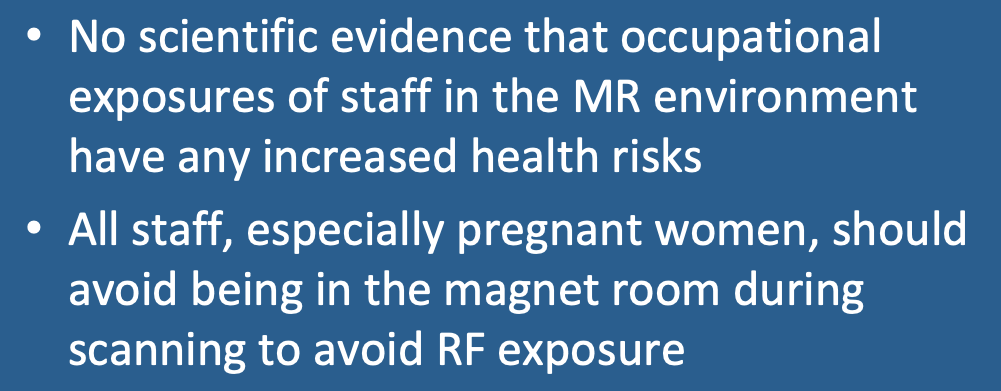
As detailed in another Q&A, there is no convincing evidence that significant long-term biological effects exist for the types of electromagnetic field exposures a staff member would be expected to have in and around an MRI scanner. The typical MRI worker is not commonly in the magnet room during scanning; therefore she is only occasionally exposed to gradient or radiofrequency fields. The main occupational exposure is from being around the static (Bo) fringe field intermittently for most of the day. With self-shielded scanners, the peak static field exposure for staff is unlikely to exceed 50% of Bo, and the average exposure over a shift would be only a few mT. 
For MR staff working around scanners with field strengths of 3T or higher, however, short term sensory effects (including nausea and dizziness) have been clearly demonstrated. For example, the incidence of vertigo among staff working at 1.5T, 3.0T, and 7.0T has been reported to be about 4%, 8%, and 24% respectively. These effects can be mitigated by moving slowly around the scanner or at the scanner bore to minimize dB/dt in the fringe field. 
Regulatory Limits
There is no shortage of regulatory organizations that have developed and promulgated standards for occupational magnetic field exposures, although most are not binding around the world. The most important organizations include the International Electrotechnical Commission (IEC), the International Commission on Non-Ionizing Radiation Protection (ICNIRP), and the Institute of Electrical and Electronics Engineers (IEEE).  In 2013, the European Council published Directive 2013/35/EU, a sweeping revision of prior regulations on occupational electromagnetic field exposure by which European Union member states are now bound. The EU Directive for the most part mirrors the exposure limits set by the ICNIRP in 2010, but have exceptions for special clinical, research, or engineering situations.
The existing panoply of regulations is confusing and in some cases contradictory. For example, the occupational body static field limit is set at 8T by the IEC, but only 2T by the ICNIRP. For occupational RF exposures, the current limits are 4 W/kg for the IEC, but only 0.4 W/kg for both the ICNIRP and IEEE. The excellent reviews by McRobbie (2012) and Mazar (2016) nicely catalog various occupational limits in various countries and their rationales.
Special Considerations for Pregnant Staff
In 1993 Kanal et al reviewed medical history questionnaires from over 1400 pregnant MRI technologists, nurses and health care workers.  They found no statistically significant difference between the rates of spontaneous abortions/miscarriages, or low birth weights, or premature delivery, or infertility, or male offspring between those working as MR employees versus the same population when they were employed elsewhere.        

Although there is no definitive evidence that occupational exposure to MR-level magnetic fields may be harmful to a pregnant staff member or her fetus, we have adopted a conservative approach and try to limit exposure throughout pregnancy.  We try to keep pregnant personnel out of the scanner rooms especially while scanning is occurring.  This practice minimizes their exposure to the higher level of static magnetic fields and eliminates completely their exposure to the time-varying (RF) fields. Being outside the room during scanning also nullifies concerns about acoustic noise on the fetus. If a staff member has strong fears about her occupational exposure during pregnancy, we assign her a clerical position far away from the scanner (a job we refer to as "tech-retary").  Fortunately, there are also other jobs in the MR suite, such as patient preparation and counseling, image archiving and filming, with little or no magnetic field exposure.

Advanced Discussion (show/hide)»

No supplementary material yet. Check back soon!

lReferences
     Directive 2013/35/EU of the European Parliament and of the Council of 26 June 2013 on the minimum health and safety requirements regarding the exposure of workers to the risks arising from physical agents (electromagnetic fields); 2013 (accessed July 2020)
     Frankel J, Wilén J, Mild KH. Assessing exposures to magnetic resonance imaging’s complex mixture of magnetic fields for in vivo, in vitro, and epidemiological studies of health effects for staff and patients. Front Public Health 2018; 6:66. [DOI Link]  
     Health Canada. Safety Code 6. Limits of human exposure to radio frequency electromagnetic energy in the frequency range from 3 kHz to 300 GHz. 2015. (accessed July 2020)
     IEEE International Committee on Electromagnetic Safety Technical Committee. Synopsis of IEEE Std C95.1™-2019 “IEEE standard for safety levels with respect to human exposure to electric, magnetic, and electromagnetic fields, 0 Hz to 300 GHz”. [DOI Link] 
     International Commission on Non-Ionizing Radiation Protection. ICNIRP Guidelines on limits of exposure to static magnetic fields. Health Phys 2009; 96:504-514. [DOI Link]
     International Commission on Non-Ionizing Radiation Protection. ICNIRP Guidelines for limiting exposure to electromagnetic fields (100 kHz to 300 GHZ). Health Phys 2020; 118:483-524. [DOI Link]
     International Electrotechnical Commission. IEC 60601-2-33:2010: Medical Electrical Equipment - Part 2-33: Particular Requirements for the Basic Safety and Essential Performance of Magnetic Resonance Equipment for Medical Diagnosis. 3rd ed. with amendments. International Electrotechnical Commission; 2015. (accessed July 2020)     
     Kanal E, Gillen J, Evans JA, et al. Survey of reproductive health among female MR workers. Radiology 1993; 187:395‑399. [DOI Link]
     Mazar H. Human radio frequency exposure limits: an update of reference levels in Europe, USA, Canada, China, Japan and Korea. IEEE International Symposium on Electromagnetic Compatibility 2016: 467-473. [DOI Link]
     McRobbie DW. Occupational exposure in MRI. Br J Radiol 2012; 85:293-312.  [DOI Link] (excellent review, now somewhat out of date, but multiple tables show the relatively diverse and complex criteria for magnetic field exposures of various nationa/international organizations). 
    Schaap K, Christopher-De Vries Y, Crozier S, et al. Exposure to static and time-varying magnetic fields from workin in the static magnetic stray fields of MRI scanners: a comprehensive survey in the Netherlands. Ann Occup Hyg 2014; 58:1094-1110. [DOI Link]
     Schaap K, Christopher-De Vries Y, Mason CK, et al. Occupational exposure of healthcare and research staff to static magnetic stray fields from 1.5-7 tesla MRI scanners is associated with transient symptoms. Occup Environ Med 2014; 71:423-429. [DOI Link]

Related Questions
     How do radiofrequency fields affect biological tissues? 
     Does MRI pose any risk to the developing fetus?  Do you scan pregnant patients?
    Who regulates/sets standards for MRI equipment and safety?  

←  Previous Question
Next Question  →
↑ Complete List of Questions ↑
© 2024 AD Elster, ELSTER LLC
All rights reserved.   
MRIquestions.com - Home
Donate
Please help keep this site free for everyone in the world!