Questions and Answers ​in MRI
  • Home
  • Complete List of Questions
  • …Magnets & Scanners
    • Basic Electromagnetism >
      • What causes magnetism?
      • What is a Tesla?
      • Who was Tesla?
      • What is a Gauss?
      • How strong is 3.0T?
      • What is a gradient?
      • Aren't gradients coils?
      • What is susceptibility?
      • How to levitate a frog?
      • What is ferromagnetism?
      • Superparamagnetism?
    • Magnets - Part I >
      • Types of magnets?
      • Brands of scanners?
      • Which way does field point?
      • Which is the north pole?
      • Low v mid v high field?
      • Advantages to low-field?
      • Disadvantages?
      • What is homogeneity?
      • Why homogeneity?
      • Why shimming?
      • Passive shimming?
      • Active shimming?
    • Magnets - Part II >
      • Superconductivity?
      • Perpetual motion?
      • How to ramp?
      • Superconductive design?
      • Room Temp supercon?
      • Liquid helium use?
      • What is a quench?
      • Is field ever turned off?
      • Emergency stop button?
    • Gradients >
      • Gradient coils?
      • How do z-gradients work?
      • X- and Y- gradients?
      • Open scanner gradients?
      • Eddy current problems?
      • Active shielded gradients?
      • Active shield confusion?
      • What is pre-emphasis?
      • Gradient heating?
      • Gradient specifications?
      • Gradient linearity?
    • RF & Coils >
      • Many kinds of coils?
      • Radiofrequency waves?
      • Phase v frequency?
      • RF Coil function(s)?
      • RF-transmit coils?
      • LP vs CP (Quadrature)?
      • Multi-transmit RF?
      • Receive-only coils?
      • Array coils?
      • AIR Coils?
    • Site Planning >
      • MR system layout?
      • What are fringe fields?
      • How to reduce fringe?
      • Magnetic shielding?
      • Need for vibration testing?
      • What's that noise?
      • Why RF Shielding?
      • Wires/tubes thru wall?
  • ...Safety and Screening
    • Overview >
      • ACR Safety Zones?
      • MR safety screening?
      • Incomplete screening?
      • Passive v active implants?
      • Conditional implants?
      • Common safety issues?
      • Projectiles?
      • Metal detectors?
      • Pregnant patients?
      • Postop, ER & ICU patients?
      • Temperature monitoring?
      • Orbital foreign bodies?
      • Bullets and shrapnel?
    • Static Fields >
      • "Dangerous" metals?
      • "Safe" metals?
      • Magnetizing metal?
      • Object shape?
      • Forces on metal?
      • Most dangerous place?
      • Force/torque testing?
      • Static field bioeffects?
      • Dizziness/Vertigo?
      • Flickering lights?
      • Metallic taste?
    • RF Fields >
      • RF safety overview?
      • RF biological effects?
      • What is SAR?
      • SAR limits?
      • Operating modes?
      • How to reduce SAR?
      • RF burns?
      • Estimate implant heating?
      • SED vs SAR?
      • B1+rms vs SAR?
      • Personnel exposure?
      • Cell phones?
    • Gradient Fields >
      • Gradient safety overview
      • Acoustic noise?
      • Nerve stimulation?
      • Gradient vs RF heating?
    • Safety: Neurological >
      • Aneurysm coils/clips?
      • Shunts/drains?
      • Pressure monitors/bolts?
      • Deep brain stimulators?
      • Spinal cord stimulators?
      • Vagal nerve stimulators?
      • Cranial electrodes?
      • Carotid clamps?
      • Peripheral stimulators?
      • Epidural catheters?
    • Safety: Head & Neck >
      • Additional orbit safety?
      • Cochlear Implants?
      • Bone conduction implants?
      • Other ear implants?
      • Dental/facial implants?
      • ET tubes & airways?
    • Safety: Chest & Vascular >
      • Breast tissue expanders?
      • Breast biopsy markers?
      • Airway stents/valves/coils?
      • Respiratory stimulators?
      • Ports/vascular access?
      • Swan-Ganz catheters?
      • IVC filters?
      • Implanted infusion pumps?
      • Insulin pumps & CGMs?
      • Vascular stents/grafts?
      • Sternal wires/implants?
    • Safety: Cardiac >
      • Pacemaker dangers?
      • Pacemaker terminology?
      • New/'Safe" Pacemakers?
      • Old/Legacy Pacemakers?
      • Violating the conditions?
      • Epicardial pacers/leads?
      • Cardiac monitors?
      • Heart valves?
      • Miscellaneous CV devices?
    • Safety: Abdominal >
      • PIllCam and capsules?
      • Gastric pacemakers?
      • Other GI devices?
      • Contraceptive devices?
      • Foley catheters?
      • Incontinence devices?
      • Penile Implants?
      • Sacral nerve stimulators?
      • GU stents and other?
    • Safety: Orthopedic >
      • Orthopedic hardware?
      • External fixators?
      • Traction and halos?
      • Bone stimulators?
      • Magnetic rods?
  • …The NMR Phenomenon
    • Spin >
      • What is spin?
      • Why I = ½, 1, etc?
      • Proton = nucleus = spin?
      • Predict nuclear spin (I)?
      • Magnetic dipole moment?
      • Gyromagnetic ratio (γ)?
      • "Spin" vs "Spin state"?
      • Energy splitting?
      • Fall to lowest state?
      • Quantum "reality"?
    • Precession >
      • Why precession?
      • Who was Larmor?
      • Energy for precession?
      • Chemical shift?
      • Net magnetization (M)?
      • Does M instantly appear?
      • Does M also precess?
      • Does precession = NMR?
    • Resonance >
      • MR vs MRI vs NMR?
      • Who discovered NMR?
      • How does B1 tip M?
      • Why at Larmor frequency?
      • What is flip angle?
      • Spins precess after 180°?
      • Phase coherence?
      • Release of RF energy?
      • Rotating frame?
      • Off-resonance?
      • Adiabatic excitation?
      • Adiabatic pulses?
    • Relaxation - Physics >
      • Bloch equations?
      • What is T1?
      • What is T2?
      • Relaxation rate vs time?
      • Why is T1 > T2?
      • T2 vs T2*?
      • Causes of Relaxation?
      • Dipole-dipole interactions?
      • Chemical Exchange?
      • Spin-Spin interactions?
      • Macromolecule effects?
      • Which H's produce signal?
      • "Invisible" protons?
      • Magnetization Transfer?
      • Bo effect on T1 & T2?
      • How to predict T1 & T2?
    • Relaxation - Clincial >
      • T1 bright? - fat
      • T1 bright? - other oils
      • T1 bright? - cholesterol
      • T1 bright? - calcifications
      • T1 bright? - meconium
      • T1 bright? - melanin
      • T1 bright? - protein/mucin
      • T1 bright? - myelin
      • Magic angle?
      • MT Imaging/Contrast?
  • …Pulse Sequences
    • MR Signals >
      • Origin of MR signal?
      • Free Induction Decay?
      • Gradient echo?
      • TR and TE?
      • Spin echo?
      • 90°-90° Hahn Echo?
      • Stimulated echoes?
      • STEs for imaging?
      • 4 or more RF-pulses?
      • Partial flip angles?
      • How is signal higher?
      • Optimal flip angle?
    • Spin Echo >
      • SE vs Multi-SE vs FSE?
      • Image contrast: TR/TE?
      • Opposite effects ↑T1 ↑T2?
      • Meaning of weighting?
      • Does SE correct for T2?
      • Effect of 180° on Mz?
      • Direction of 180° pulse?
    • Inversion Recovery >
      • What is IR?
      • Why use IR?
      • Phase-sensitive IR?
      • Why not PSIR always?
      • Choice of IR parameters?
      • TI to null a tissue?
      • STIR?
      • T1-FLAIR
      • T2-FLAIR?
      • IR-prepped sequences?
      • Double IR?
    • Gradient Echo >
      • GRE vs SE?
      • Multi-echo GRE?
      • Types of GRE sequences?
      • Commercial Acronyms?
      • Spoiling - what and how?
      • Spoiled-GRE parameters?
      • Spoiled for T1W only?
      • What is SSFP?
      • GRASS/FISP: how?
      • GRASS/FISP: parameters?
      • GRASS vs MPGR?
      • PSIF vs FISP?
      • True FISP/FIESTA?
      • FIESTA v FIESTA-C?
      • DESS?
      • MERGE/MEDIC?
      • GRASE?
      • MP-RAGE v MR2RAGE?
    • Susceptibility Imaging >
      • What is susceptibility (χ)?
      • What's wrong with GRE?
      • Making an SW image?
      • Phase of blood v Ca++?
      • Quantitative susceptibility?
    • Diffusion: Basic >
      • What is diffusion?
      • Iso-/Anisotropic diffusion?
      • "Apparent" diffusion?
      • Making a DW image?
      • What is the b-value?
      • b0 vs b50?
      • Trace vs ADC map?
      • Light/dark reversal?
      • T2 "shine through"?
      • Exponential ADC?
      • T2 "black-out"?
      • DWI bright causes?
    • Diffusion: Advanced >
      • Diffusion Tensor?
      • DTI (tensor imaging)?
      • Whole body DWI?
      • Readout-segmented DWI?
      • Small FOV DWI?
      • IVIM?
      • Diffusion Kurtosis?
    • Fat-Water Imaging >
      • Fat & Water properties?
      • F-W chemical shift?
      • In-phase/out-of-phase?
      • Best method?
      • Dixon method?
      • "Fat-sat" pulses?
      • Water excitation?
      • STIR?
      • SPIR?
      • SPAIR v SPIR?
      • SPIR/SPAIR v STIR?
  • …Making an Image
    • From Signals to Images >
      • Phase v frequency?
      • Angular frequency (ω)?
      • Signal squiggles?
      • Real v Imaginary?
      • Fourier Transform (FT)?
      • What are 2D- & 3D-FTs?
      • Who invented MRI?
      • How to locate signals?
    • Frequency Encoding >
      • Frequency encoding?
      • Receiver bandwidth?
      • Narrow bandwidth?
      • Slice-selective excitation?
      • SS gradient lobes?
      • Cross-talk?
      • Frequency encode all?
      • Mixing of slices?
      • Two slices at once?
      • Simultaneous Multi-Slice?
    • Phase Encoding >
      • Phase-encoding gradient?
      • Single PE step?
      • What is phase-encoding?
      • PE and FE together?
      • 2DFT reconstruction?
      • Choosing PE/FE direction?
    • Performing an MR Scan >
      • What are the steps?
      • Automatic prescan?
      • Routine shimming?
      • Coil tuning/matching?
      • Center frequency?
      • Transmitter gain?
      • Receiver gain?
      • Dummy cycles?
      • Where's my data?
      • MR Tech qualifications?
    • Image Quality Control >
      • Who regulates MRI?
      • Who accredits?
      • Mandatory accreditation?
      • Routine quality control?
      • MR phantoms?
      • Geometric accuracy?
      • Image uniformity?
      • Slice parameters?
      • Image resolution?
      • Signal-to-noise?
      • Ghosting?
  • …K-space & Rapid Imaging
    • K-space (Basic) >
      • What is k-space?
      • Parts of k-space?
      • What does "k" stand for?
      • Spatial frequencies?
      • Locations in k-space?
      • Data for k-space?
      • Why signal ↔ k-space?
      • Spin-warp imaging?
      • Big spot in middle?
      • K-space trajectories?
      • Radial sampling?
    • K-space (Advanced) >
      • K-space grid?
      • Negative frequencies?
      • Field-of-view (FOV)
      • Rectangular FOV?
      • Partial Fourier?
      • Phase symmetry?
      • Read symmetry?
      • Why not use both?
      • ZIP?
    • Rapid Imaging (FSE &EPI) >
      • What is FSE/TSE?
      • FSE parameters?
      • Bright Fat?
      • Other FSE differences?
      • Dual-echo FSE?
      • Driven equilibrium?
      • Reduced flip angle FSE?
      • Hyperechoes?
      • SPACE/CUBE/VISTA?
      • Echo-planar imaging?
      • HASTE/SS-FSE?
    • Parallel Imaging (PI) >
      • What is PI?
      • How is PI different?
      • PI coils and sequences?
      • Why and when to use?
      • Two types of PI?
      • SENSE/ASSET?
      • GRAPPA/ARC?
      • CAIPIRINHA?
      • Compressed sensing?
      • Noise in PI?
      • Artifacts in PI?
  • …Contrast Agents
    • Contrast Agents: Physics >
      • Why Gadolinium?
      • Paramagnetic relaxation?
      • What is relaxivity?
      • Why does Gd shorten T1?
      • Does Gd affect T2?
      • Gd & field strength?
      • Best T1-pulse sequence?
      • Triple dose and MT?
      • Dynamic CE imaging?
      • Gadolinium on CT?
    • Contrast Agents: Clinical >
      • So many Gd agents!
      • Important properties?
      • Ionic v non-ionic?
      • Intra-articular/thecal Gd?
      • Gd liver agents (Eovist)?
      • Mn agents (Teslascan)?
      • Feridex & Liver Agents?
      • Lymph node agents?
      • Ferumoxytol?
      • Blood pool (Ablavar)?
      • Bowel contrast agents?
    • Contrast Agents: Safety >
      • Gadolinium safety?
      • Allergic reactions?
      • Renal toxicity?
      • What is NSF?
      • NSF by agent?
      • Informed consent for Gd?
      • Gd protocol?
      • Is Gd safe in infants?
      • Reduced dose in infants?
      • Gd in breast milk?
      • Gd in pregnancy?
      • Gd accumulation?
      • Gd deposition disease?
  • …Cardiovascular and MRA
    • Flow effects in MRI >
      • Defining flow?
      • Expected velocities?
      • Laminar v turbulent?
      • Predicting MR of flow?
      • Time-of-flight effects?
      • Spin phase effects?
      • Flow void?
      • Why GRE ↑ flow signal?
      • Slow flow v thrombus?
      • Even-echo rephasing?
      • Flow-compensation?
      • Flow misregistration?
    • MR Angiography - I >
      • MRA methods?
      • Dark vs bright blood?
      • Time-of-Flight (TOF) MRA?
      • 2D vs 3D MRA?
      • MRA parameters?
      • Magnetization Transfer?
      • Ramped flip angle?
      • MOTSA?
      • Fat-suppressed MRA?
      • TOF MRA Artifacts?
      • Phase-contrast MRA?
      • What is VENC?
      • Measuring flow?
      • 4D Flow Imaging?
      • How accurate?
    • MR Angiography - II >
      • Gated 3D FSE MRA?
      • 3D FSE MRA parameters?
      • SSFP MRA?
      • Inflow-enhanced SSFP?
      • MRA with ASL?
      • Other MRA methods?
      • Contrast-enhanced MRA?
      • Timing the bolus?
      • View ordering in MRA?
      • Bolus chasing?
      • TRICKS or TWIST?
      • CE-MRA artifacts?
    • Cardiac I - Intro/Anatomy >
      • Cardiac protocols?
      • Patient prep?
      • EKG problems?
      • Magnet changes EKG?
      • Gating v triggering?
      • Gating parameters?
      • Heart navigators?
      • Dark blood/Double IR?
      • Why not single IR?
      • Triple IR?
      • Polar plots?
      • Coronary artery MRA?
    • Cardiac II - Function >
      • Beating heart movies?
      • Cine parameters?
      • Real-time cine?
      • Ventricular function?
      • Tagging/SPAMM?
      • Perfusion: why and how?
      • 1st pass perfusion?
      • Quantifying perfusion?
      • Dark rim artifact
    • Cardiac III - Viability >
      • Gd enhancement?
      • TI to null myocardium?
      • PS (phase-sensitive) IR?
      • Wideband LGE?
      • T1 mapping?
      • Iron/T2*-mapping?
      • Edema/T2-mapping?
      • Why/how stress test?
      • Stess drugs/agents?
      • Stress consent form?
  • …MR Artifacts
    • Tissue-related artifacts >
      • Chemical shift artifact?
      • Chemical shift in phase?
      • Reducing chemical shift?
      • Chemical Shift 2nd Kind?
      • In-phase/out-of phase?
      • IR bounce point?
      • Susceptibility artifact?
      • Metal suppression?
      • Dielectric effect?
      • Dielectric Pads?
    • Motion-related artifacts >
      • Why discrete ghosts?
      • Motion artifact direction?
      • Reducing motion artifacts?
      • Saturation pulses?
      • Gating methods?
      • Respiratory comp?
      • Navigator echoes?
      • PROPELLER/BLADE?
    • Technique-related artifacts >
      • Partial volume effects?
      • Slice overlap?
      • Aliasing?
      • Wrap-around artifact?
      • Eliminate wrap-around?
      • Phase oversampling?
      • Frequency wrap-around?
      • Spiral/radial artifacts?
      • Gibbs artifact?
      • Nyquist (N/2) ghosts?
      • Zipper artifact?
      • Data artifacts?
      • Surface coil flare?
      • MRA Artifacts (TOF)?
      • MRA artifacts (CE)?
  • …Functional Imaging
    • Perfusion I: Intro & DSC >
      • Measuring perfusion?
      • Meaning of CBF, MTT etc?
      • DSC v DCE v ASL?
      • How to perform DSC?
      • Bolus Gd effect?
      • T1 effects on DSC?
      • DSC recirculation?
      • DSC curve analysis?
      • DSC signal v [Gd]
      • Arterial input (AIF)?
      • Quantitative DSC?
    • Perfusion II: DCE >
      • What is DCE?
      • How is DCE performed?
      • How is DCE analyzed?
      • Breast DCE?
      • DCE signal v [Gd]
      • DCE tissue parmeters?
      • Parameters to images?
      • K-trans = permeability?
      • Utility of DCE?
    • Perfusion III: ASL >
      • What is ASL?
      • ASL methods overview?
      • CASL?
      • PASL?
      • pCASL?
      • ASL parameters?
      • ASL artifacts?
      • Gadolinium and ASL?
      • Vascular color maps?
      • Quantifying flow?
    • Functional MRI/BOLD - I >
      • Who invented fMRI?
      • How does fMRI work?
      • BOLD contrast?
      • Why does BOLD ↑ signal?
      • Does BOLD=brain activity?
      • BOLD pulse sequences?
      • fMRI Paradigm design?
      • Why "on-off" comparison?
      • Motor paradigms?
      • Visual?
      • Language?
    • Functional MRI/BOLD - II >
      • Process/analyze fMRI?
      • Best fMRI software?
      • Data pre-processing?
      • Registration/normalization?
      • fMRI statistical analysis?
      • General Linear Model?
      • Activation "blobs"?
      • False activation?
      • Resting state fMRI?
      • Analyze RS-fMRI?
      • Network/Graphs?
      • fMRI at 7T?
      • Mind reading/Lie detector?
      • fMRI critique?
  • …MR Spectroscopy
    • MRS I - Basics >
      • MRI vs MRS?
      • Spectra vs images?
      • Chemical shift (δ)?
      • Measuring δ?
      • Backward δ scale?
      • Predicting δ?
      • Size/shapes of peaks?
      • Splitting of peaks?
      • Localization methods?
      • Single v multi-voxel?
      • PRESS?
      • STEAM?
      • ISIS?
      • CSI?
    • MRS II - Clinical ¹H MRS >
      • How-to: brain MRS?
      • Water suppression?
      • Fat suppression?
      • Normal brain spectra?
      • Choice of TR/TE/etc?
      • Hunter's angle?
      • Lactate inversion?
      • Metabolite mapping?
      • Metabolite quantitation?
      • Breast MRS?
      • Gd effect on MRS?
      • How-to: prostate MRS?
      • Prostate spectra?
      • Muscle ¹H-MRS?
      • Liver ¹H-MRS?
      • MRS artifacts?
    • MRS III - Multi-nuclear >
      • Other nuclei?
      • Why phosphorus?
      • How-to: ³¹P MRS
      • Normal ³¹P spectra?
      • Organ differences?
      • ³¹P measurements?
      • Decoupling?
      • NOE?
      • Carbon MRS?
      • Sodium imaging?
      • Xenon imaging?
  • ...Artificial Intelligence
    • AI Part I: Basics >
      • Artificial Intelligence (AI)?
      • What is a neural network?
      • Machine Learning (ML)?
      • Shallow v Deep ML?
      • Shallow networks?
      • Deep network types?
      • Data prep and fitting?
      • Back-Propagation?
      • DL 'Playground'?
    • AI Part 2: Advanced >
      • What is convolution?
      • Convolutional Network?
      • Softmax?
      • Upsampling?
      • Limitations/Problems of AI?
      • Is the Singularity near?
    • AI Part 3: Image processing >
      • AI in clinical MRI?
      • Super-resolution?
  • ...Tissue Properties Imaging
    • MRI of Hemorrhage >
      • Hematoma overview?
      • Types of Hemoglobin?
      • Hyperacute/Oxy-Hb?
      • Acute/Deoxy-Hb?
      • Subacute/Met-Hb?
      • Deoxy-Hb v Met-Hb?
      • Extracellular met-Hb?
      • Chronic hematomas?
      • Hemichromes?
      • Ferritin/Hemosiderin?
      • Subarachnoid blood?
      • Blood at lower fields?
    • T2 cartilage mapping
    • MR Elastography?
    • Synthetic MRI?
    • Amide Proton Transfer?
    • MR thermography?
    • Electric Properties Imaging?
  • Copyright/Legal
    • Copyright Issues
    • Legal Disclaimers
  • Forums/Blogs/Links
  • What's New
  • Self-test Quizzes - NEW!
    • Magnets & Scanners Quiz
    • Safety & Screening Quiz
    • NMR Phenomenon Quiz
    • Pulse Sequences Quiz
    • Making an Image Quiz
    • K-space & Rapid Quiz
    • Contrast & Blood Quiz
    • Cardiovascular & MRA Quiz

Myocardial T1-Mapping

How is cardiac T1 mapping performed? When is it useful?  
myocardial T1 mapping
Many diffuse diseases of heart muscle (cadiomyopathies) may not produce focal changes in signal intensity, motion, or enhancement on routine cardiac MRI studies. These disorders, however, may affect global myocardial NMR properties, such as T1, T2, or T2* relaxation times. Specialized pulse sequences optimized for cardiac imaging are able to estimate these relaxation values and even create color anatomic overlay maps for each parameter. On this web page I will discuss T1-mapping; later Q&A's will describe T2-mapping (for edema detection) and T2* measurements (for iron deposition).
cardiac T1 map
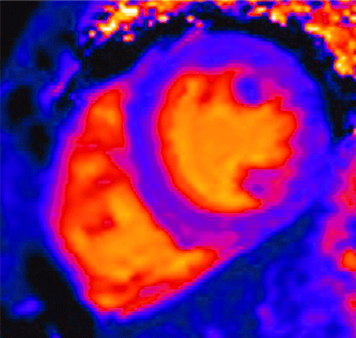
Colored T1 map from which T1 values can be measured.
The T1 value of normal myocardium at 1.5T is approximately 950-1000 ms at 1.5T and 1100-1250 at 3.0T. Myocardial T1 is prolonged in most forms of pathology, including edema (from acute infarction or myocarditis) and as the end result of many chronic cardiac and systemic disorders (such as hypertrophic, toxic, and dilated cardiomyopathies; remote ischemia/infarction; diabetes; valvular disease; amyloidosis; and sarcoidosis). T1 shortening due to pathology is rare, but can be seen in disorders like Fabry disease, fatty arrythmogenic right ventricular dysplasia (ARVD), or hemochromatosis where glycoproteins, lipids, or iron respectively are deposited in the myocardium. Of course, gadolinium contrast accumulation shortens T1 values in both healthy and diseased myocardium.
T1-myocardial mapping may be performed either with or without contrast. When done without contrast it is commonly referred to as "native" T1-mapping. Administration of gadolinium contrast shortens the T1 of both normal and diseased myocardium. Postcontrast T1-mapping may be useful to better differentiate and quantify enhancement in various areas of the heart.
The traditional "gold standard" method for determining tissue T1 is to perform a series of independent single-point inversion recovery signal measurements at different TI's.  Very long TR's are required to allow the restoration of full longitudinal magnetization Mo between measurements. These data points lie along an exponential growth curve (shown in green in the diagram right). By mathematical fitting T1 can be easily calculated. The single-point IR method may require 20+ minutes of data sampling, and hence is too time-consuming for practical clinical application. 
Look Locker
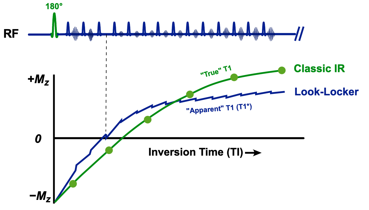
A comparison of longitudinal magnetization (Mz) using the classic (but time-consuming) single-point IR method with a rapid GRE method (Look-Locker). The latter method produces a lower equilibrium magnetization and consistently shorter T1 estimates, often denoted T1*.
Even before the development of MR imaging, laboratory NMR chemists searched for faster ways to determine the T1's of their samples. In 1970 Look and Locker reasoned that instead of making a single measurement at a fixed TI after inversion, many more data points could be acquired by sampling a periodic train of rapidly applied constant flip-angle pulses. If the RF-pulse train were sufficiently long, the longitudinal magnetization would be driven to a new steady state (which, of course, would be lower than the undisturbed equilibrium state (Mo) of the classic IR method). An estimate for T1 could nevertheless be derived by observing the rate of change of signal amplitudes sampled during this periodic pulse train.
Notwithstanding the theoretical corrections, T1 values computed by the Look-Locker method depend on a number of factors, including type of pulse sequence (GRE vs b-SSFP), flip angle, TR, tissue T2, field strength, magnetization transfer effects, RF-inhomogeneity, and heart rate. Because of this experimental dependency, we are not measuring "true" T1, but a consistently shorter "apparent" T1. Analogous to the term used in T2 relaxation, apparent T1 is often called T1* ("T1-star"). 
At present the most widely used T1 measurement sequence is Modified Look-Locker Imaging (MOLLI). The basic strategy is to use only 2-3 inversion pulses followed by several single-shot b-SSFP readouts at various fixed inversion times. Short rest periods are interspersed to allow recovery of longitudinal magnetization between cycles. In conjunction with parallel imaging and conjugate symmetry methods, MOLLI allows accurate T1-values of myocardium to be obtained in only one breath-hold per slice (~20 sec).
Picture
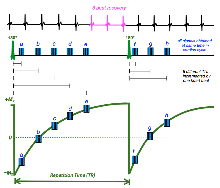
Modified Look-Locker Imaging (MOLLI) with 5(3)3 pattern.
In one common implementation illustrated above, 5(3)3 MOLLI, measurements are obtained at different TI's over 5+3=8 heart beats with a 3 beat recovery period in between. The inversion pulses are placed so that readout periods (a, b, c, ...) all occur at the same place in the cardiac cycle, allowing pixel-wise T1 calculations to be performed. The recovery period may be reduced on post-contrast imaging due to the lower T1 values of gadolinium-containing myocardium. For patients who cannot suspend respiration for 20+ seconds, shortened MOLLI (shMOLLI) using a 5-1-1 scheme is available. 
SASHA MRI
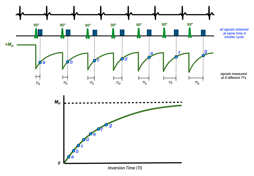
SASHA Technique of T1-mapping. Multiple 90°-pulses tip the magnetization into the transverse plane with each heart beat. Signal readouts are performed at the same position in the cardiac cycle, but at different TI's. A baseline data point (not shown) is acquired without magnetization preparation. By curve fitting T1 can be computed for each point.
Saturation recovery (SR) methods for T1 mapping are becoming increasingly popular, the best known of which is called SASHA (SAturation-recovery single-SHot Acquisition). SR recovery methods use 90°- instead of 180°-pulses, which have the advantage of removing magnetization memory from prior cycles and allowing a direct estimate of true T1 to be made. Disadvantages include lower SNR and more artifacts.

Advanced Discussion (show/hide)»

A step beyond simple T1-mapping is calculation of extracellular myocardial volume (ECV). The T1-relaxation rate (R1=1/T1) of any tissue changes proportionally with the gadolinium concentration. The T1 mapping methods described above allow the relaxation rates of blood and myocardium to be calculated before and after contrast administration. Assuming fast exchange of water molecules and a 2-compartment model, the ECV can then be calculated by the formula

ECV = {ΔR1myocardium / ΔR1blood} × {1 − Hct}

where ΔR1 is the change of relaxation rates for myocardium and blood pre- and post-contrast, and Hct is the hematocrit. (An adjustment for Hct must be made since gadolinium resides only in plasma and does not enter intact red blood cells).


References
     Aherne E, Chow K, Carr J. Cardiac T1 mapping: techniques and applications. J Magn Reson Imaging 2020; 51:1336-1356. (excellent recent review)
     Burt JR, Zimmerman SL, Kamel IR, et al. Myocardial T1 mapping: techniques and potential applications. RadioGraphics 2014; 34:377-395. 
     Chow K, Flewitt JA, Green JD, et al. Saturation recovery single-shot acquisition (SASHA) for myocardial T(1) mapping. Magn Reson Med 2014; 71:2082-95.
     Haaf P, Garg P, Messroghli DR, et al. Cardiac T1 mapping and extracellular volume (ECV) in clinical practice: a comprehensive review. J Cardiovasc Magn Reson 2016; 18:69. (excellent recent review with a clinical focus)
     Hamlin SA, Henry TS, Little BP, et al. Mapping the future of cardiac MR imaging: case-based review of T1 and T2 mapping techniques. Radiographics 2014; 34:1594-1611.
     Jerosch-Herold M, Kwong RY. Cardiac T1 imaging. Top Magn Reson Imaging 2014; 23:3-11.
     Kellman P, Hansen MS. T1-mapping in the heart: accuracy and precision. J Cardiovasc Magn Reson 2014; 16:2. (great review discussing advantages and limitations of each technique)
     Look DC. Locker DR. Time saving in measurement of NMR and EPR relaxation times. Rev Sci Instrum 1970; 41:250-1. (original technique developed for chemical NMR, not practical for clinical MRI).
     Messroghli DR, Moon JC, Ferreira VM, et al. Clinical recommendations for cardiovascular magnetic resonance mapping of T1, T2, T2* and extracellular volume: a consensus statement by the Society for Cardiovascular Magnetic Resonance (SCMR) endorsed by the European Association for Cardiovascular Imaging (EACVI). J Cardiovasc Magn Reson. 2017;19:75.
     Messroghli DR, Plein S, Higgins DM, et al. Human myocardium: single-breath-hold MR T1 mapping with high spatial resolution—reproducibility study. Radiology 2006; 238:1004-1012.
     Messroghli DR, Radjenovic A, Kozerke S, et al. Modified Look-Locker Inversion Recovery (MOLLI) for high-resolution T1 mapping of the heart. Magn Reson Med 2004; 52:141-146.     
     Moon JC, Messroghli DR, Kellman P, et al. Myocardial T1 mapping and extracellular volume quantification: a Society for Cardiovascular Magnetic Resonance (SCMR) and CMR Working Group of the European Society of Cardiology consensus statement. J Cardiovasc Magn Reson 2013; 15:92.
     Piechnik SK, Ferreira VM, Dall'Armellina E, et al. Shortened modified Look-Locker inversion recovery (shMOLLI) for clinical myocardial T1-mapping at 1.5 and 3 T within a 9 heartbeat breathhold. J Cardiovasc Magn Reson 2010; 12:69
     Schelbert EB, Messroghli DR. State of the art: Clinical applications of cardiac T1 mapping. Radiology 2016; 278:658-676.

Related Questions
     How do you set the IR parameters to achieve desired image contrast?

←  Previous Question
Next Question  →
↑ Complete List of Questions ↑
© 2024 AD Elster, ELSTER LLC
All rights reserved.   
MRIquestions.com - Home
Donate
Please help keep this site free for everyone in the world!