Fat SuppressionHow and why is fat suppression performed in an MRS study?
|
|
Adipose tissue is predominantly composed of long-chain triglycerides and free fatty acids, whose prinicipal resonances lie between δ = 0.9 and 1.4 ppm. Signal from extraneous fat can "bleed into" and contaminate the spectra from within the defined volume of interest. Lactate at δ = 1.3 is especially affected.
Lipid contamination is particularly problematic in breast MRS (since the normal breast contains large amount of fat) and for prostate MRS (since the prostate is a small organ surrounded in pelvic fat). In brain MRS, scalp and marrow fat can affect the spectra from voxels obtained near the brain surface.
|
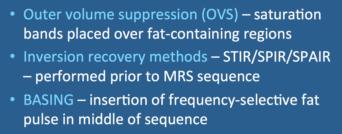
The most common method to eliminate unwanted fat signal is to place multiple saturation bands over lipid-containing regions. This method is known as outer volume suppression (OVS).
OVS bands are spatially but not frequency specific, reducing or eliminating signals from all tissues (not just lipids). They can be even thicker than the ones shown in the figure and brought down close to the edges of a single voxel even at the center of the brain, completely surrounding it. They are also commonly placed in the planes above and below the volume of interest (not illustrated). |
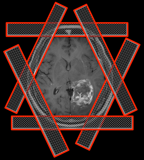
Outer volume suppression bands |
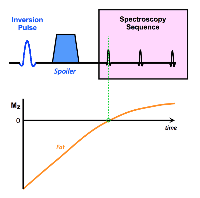 Inversion recovery (IR) method for fat suppression
OVS bands are usually sufficient for brain spectroscopy, but for body applications (especially prostate, breast and skeletal muscle MRS) additional fat suppression methods may be necessary. Inversion recovery methods widely used in conventional MR imaging may be employed, including STIR (Short TI Inversion Recovery) and SPIR (Spectral Presaturation with Inversion Recovery). A current popular choice is SPAIR (SPectral Attenuated Inversion Recovery), described more completely in a prior Q&A.
SPAIR begins with a frequency-selective, 180°-adiabatic pulse that inverts the longitudinal magnetization of fat. A time delay (~165 ms at 1.5T, ~200 ms at 3.0T) is interposed so that fat magnetization partially recovers to pass through zero at the time of spectroscopic excitation. The frequency band of the SPAIR pulse must include principal fat resonances at 0.9 and 1.3 ppm, and thus will obscure metabolites in this region of the spectrum, including lactate at δ = 1.33.
SPAIR begins with a frequency-selective, 180°-adiabatic pulse that inverts the longitudinal magnetization of fat. A time delay (~165 ms at 1.5T, ~200 ms at 3.0T) is interposed so that fat magnetization partially recovers to pass through zero at the time of spectroscopic excitation. The frequency band of the SPAIR pulse must include principal fat resonances at 0.9 and 1.3 ppm, and thus will obscure metabolites in this region of the spectrum, including lactate at δ = 1.33.

References
Felmlee JP, Ehman RL. Spatial presaturation: a method for suppressing flow artifacts and improving depiction of vascular anatomy in MR imaging. Radiology 1987;164:559 –564.
Kaldoudi E, Williams SC, Barker GJ, Tofts PS. A chemical shift selective inversion recovery sequence for fat-suppressed MRI: theory and experimental validation. Magn Reson Imaging 1993; 11:341-355.
Star-Lack J, Nelson SJ, Kurhanewicz J, et al. Improved water and lipid suppression for 3D PRESS CSI using RF band selective inversion with gradient dephasing (BASING). Magn Reson Med. 1997; 38:311–321.
Related Questions
What makes fat and water behave so differently on MRI?
There seem to be many methods available for fat suppression. Which one is the best?
What is SPAIR? How is it different than SPIR?
Why and how do you suppress water signal in MRS?
What makes fat and water behave so differently on MRI?
There seem to be many methods available for fat suppression. Which one is the best?
What is SPAIR? How is it different than SPIR?
Why and how do you suppress water signal in MRS?