TOF MRA Artifacts
What are the major TOF MR artifacts should we know about?
|
|
At least 6 different artifacts are commonly noted in time-of-flight (TOF) MRA studies. These are illustrated and briefly discussed below:
|
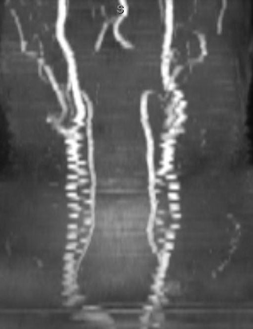
Stair-Step Artifact (2D TOF only)
This simplest form of this artifact is a subtle pixelated appearance to obliquely oriented vessels. This occurs because the slices in 2D are relatively thick (1-3 mm) compared to the in-plane spatial resolution of 0.5-1.0 mm. Stair-step artifacts can be minimized by overlapping slices by 25-30%. This means more slices are needed to cover the same area, however, with a concomitant increase in scan time.
|
Mild "stair-step" artifact due to non-isotropic voxels
|
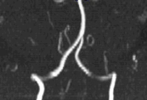
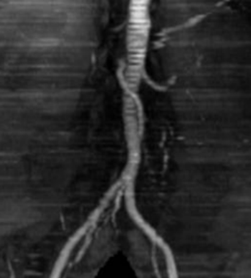
More severe stair-step artifacts occur when subtle or gross motion occurs between the individually acquried 2D slices.
Mild motion artifact causing horizontal banding on this 2D-TOF MRA of the aorta
|
Severe artifacts with jagged edges due to gross motion for carotid TOF MRA study
|
In-Plane Saturation Artifact
TOF MRA relies on the inflow of fresh (unsaturated) blood to produce high intravascular signal. This signal is highest whenever the direction of flow is perpendicular to the primary imaging plane. When vessels travel within plane, their blood may become saturated like stationary tissues, resulting in decreased signal. This may occur with either 2D or 3D TOF techniques
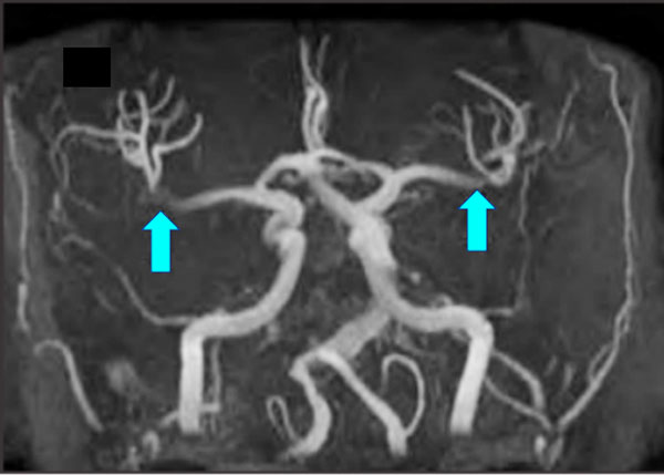
3D-TOF MRA showing artifactual in-plane signal loss within both middle cerebral arteries
|
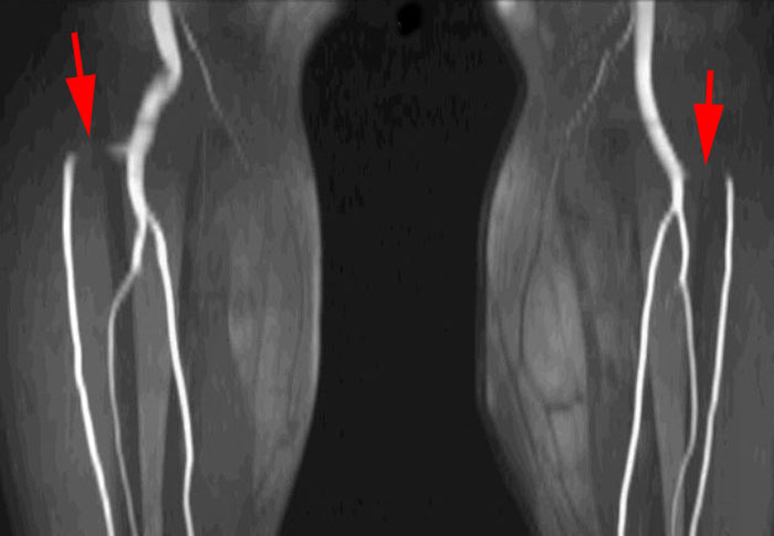
2D-TOF MRA shows artifactual in-plane signal loss in horizontal portions of both anterior tibial arteries
|
Shine-through Artifacts
|
Both 2D and 3D TOF MR angiographic images are displayed using a maximum intensity projection (MIP) algorithm. The MIP selects all pixels having an arbitrary minimum signal intensity (e.g., 2 standard deviations above background).
In addition to bright blood vessels, any other material with high signal intensity will "shine through" and "contaminate" the MIP image. Since underlying TOF MRA pulse sequences are T1-weighted, short T1 materials (hematoma, gadolinium, fat) are primarily responsible for this phenomenon. However, any high signal focus (due to motion or susceptibility) could create problems in interpretation.
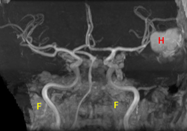
3D TOF MRA showing shine through of high signal from hematoma (H) and fat (F) at skull base
|
CSF inflow phenomenon (on source image below) creates artifact on MIP image (above)
|
Flow-Reversal Artifact
This is more of a pitfall rather than an artifact, but an important one of which to be aware. Recall that TOF techniques typically employ traveling saturation pulses to eliminate signal from veins flowing in the opposite direction. But what happens if an artery has retrograde flow due to some abnormal condition? Yes, it will be suppressed as well and not seen on the TOF MRA. An example is shown below of a patient with subclavian steal syndrome that causes flow in the left vertebral artery to be reversed. Similar phenomena may be seen in the pelvis and lower extremities where collaterals around stenotic lesions may follow a retrograde course.
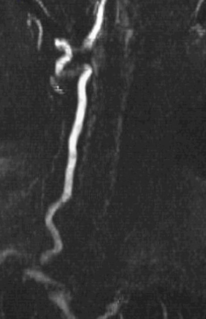
2D TOF MRA shows only right vertebral artery. No flow related signal in the left vertebral artery is seen.
|
Contrast-enhanced MRA shows retrograde filling of left vertebral artery (subclavian steal phenomenon)
|
|
Venetian Blind Artifact (3D MRA only)
This is a specific artifact seen only with the MOTSA (Multiple Overlapping Thin Slab Acquisition) technique. It represents overlap between adjacent slabs that are acquired as separate acquisitions and then fused together. The MOTSA technique is described in more detail in another Q&A.
|
|
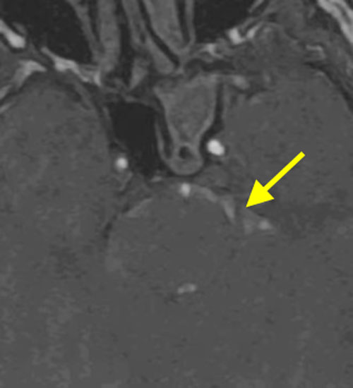
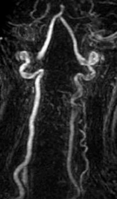
Susceptibility Artifacts
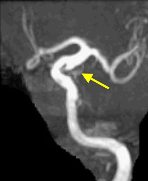
Susceptibility refers to the magnetic field distortion that occurs especially at air-tissue interfaces and near metallic objects. These distortions create magnetic gradients that cause accelerated dephasing of spins, signal loss, and spatial distortion. In MRA susceptibility artifacts are commonly encountered around surgical clips, endovascular coils, and stents. When mild, these artifacts may suggest vascular narrowing or stenosis when none exists. When severe they may cause complete loss of flow-related enhancement falsely suggesting occlusion.
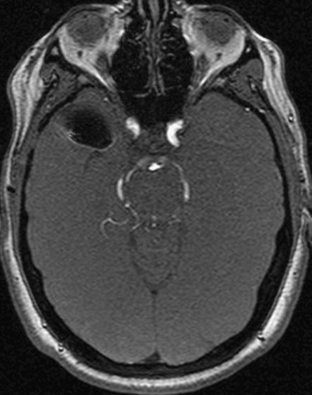
Source image shows susceptibility field distortion due to aneurysm clip
|
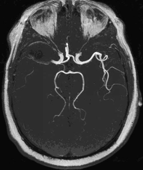
MRA show spurious loss of flow in entire right middle cerebral artery territory due to susceptibility artifact from clip
|
References
Kaufman JA, McCarter D, Geller SC, Waltman AC. Two-dimensional time-of-flight MR angiography of the lower extremities: artifacts and pitfalls. AJR Am J Roentgenol 1998; 171:129-135.
Kaufman JA, McCarter D, Geller SC, Waltman AC. Two-dimensional time-of-flight MR angiography of the lower extremities: artifacts and pitfalls. AJR Am J Roentgenol 1998; 171:129-135.
Related Questions
How does time-of-flight MRA work?
How does time-of-flight MRA work?