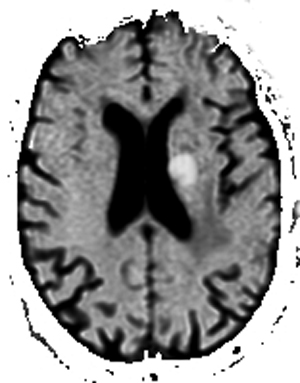
Exponential ADC
How does the "exponential" ADC map differ from a "conventional" ADC map?
|
|
As discussed in the last Q&A, lesions bright on diffusion-weighted (DW) images do not all restrict diffusion. Some have prolonged T2 values that spill over into the DW image, a phenomenon known as T2-"shine through". To sort this out, the ADC map is typically consulted. However, the ADC map has a gray scale opposite to the DW image and this may lead to some confusion.
To overcome the need to look at two separate diffusion image sets, in 1998 Jim Provenzale and colleagues proposed using an exponential image, which was simply the trace DW image divided by the b0 image. Algebraically
To overcome the need to look at two separate diffusion image sets, in 1998 Jim Provenzale and colleagues proposed using an exponential image, which was simply the trace DW image divided by the b0 image. Algebraically

where So and SDWI are the signal intensities of the b0 and conventional DW images at a given point. An example using Trace DWI, ADC. and Exponential images is shown below.
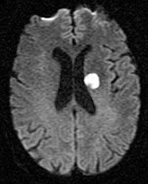
Trace DWI
|
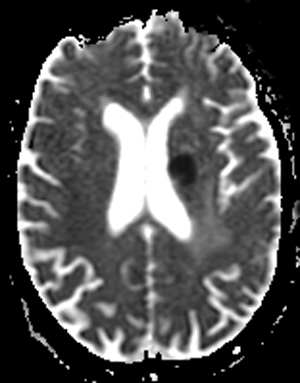
Conventional ADC
|
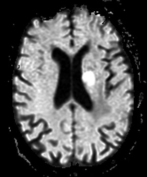
Exponential ADC
|
Other names given to the exponential image include the exponential ADC map, the exponential diffusion-weighted image, the attenuation coefficient (AC) map, and the attenuation factor map.
|
The automatic generation of exponential diffusion images is now possible on many brands of scanners. I personally don't use exponential images routinely (except for brain MRI in neonates and young infants where I find it helpful because of the intrinsically high water content and T2 signal in this age group). Because the exponential image is a calculated map, like the ADC map it is often of lower quality than the DW image. So I feel I cannot give up inspecting the DW image especially when searching for small lesions like brainstem infarctions where the additive contrast effects of prolonged T2 plus restricted diffusion are helpful. If you don't have an exponential image available, a reasonable alternative is to simply invert the conventional ADC map on your monitor.
|
Inverted conventional ADC
|
References
Provenzale JM, Engelter ST, Petrella JR, et al. Use of MR exponential diffusion-weighted images to eradicate T2 “shine-through” effect. AJR Amer J Roentgenol 1998; 172:537-539.
Provenzale JM, Engelter ST, Petrella JR, et al. Use of MR exponential diffusion-weighted images to eradicate T2 “shine-through” effect. AJR Amer J Roentgenol 1998; 172:537-539.