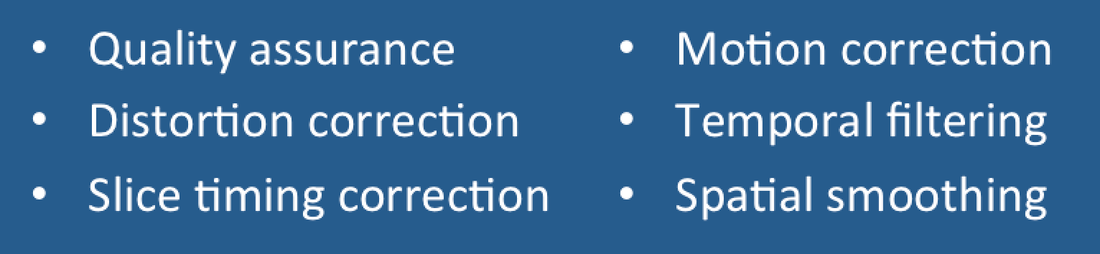
Questions and Answers ​in MRI
  • Home
  • Complete List of Questions
  • …Magnets & Scanners
    • Basic Electromagnetism >
      • What causes magnetism?
      • What is a Tesla?
      • Who was Tesla?
      • What is a Gauss?
      • How strong is 3.0T?
      • What is a gradient?
      • Aren't gradients coils?
      • What is susceptibility?
      • How to levitate a frog?
      • What is ferromagnetism?
      • Superparamagnetism?
    • Magnets - Part I >
      • Types of magnets?
      • Brands of scanners?
      • Which way does field point?
      • Which is the north pole?
      • Low v mid v high field?
      • Advantages to low-field?
      • Disadvantages?
      • What is homogeneity?
      • Why homogeneity?
      • Why shimming?
      • Passive shimming?
      • Active shimming?
    • Magnets - Part II >
      • Superconductivity?
      • Perpetual motion?
      • How to ramp?
      • Superconductive design?
      • Room Temp supercon?
      • Liquid helium use?
      • What is a quench?
      • Is field ever turned off?
      • Emergency stop button?
    • Gradients >
      • Gradient coils?
      • How do z-gradients work?
      • X- and Y- gradients?
      • Open scanner gradients?
      • Eddy current problems?
      • Active shielded gradients?
      • Active shield confusion?
      • What is pre-emphasis?
      • Gradient heating?
      • Gradient specifications?
      • Gradient linearity?
    • RF & Coils >
      • Many kinds of coils?
      • Radiofrequency waves?
      • Phase v frequency?
      • RF Coil function(s)?
      • RF-transmit coils?
      • LP vs CP (Quadrature)?
      • Multi-transmit RF?
      • Receive-only coils?
      • Array coils?
      • AIR Coils?
    • Site Planning >
      • MR system layout?
      • What are fringe fields?
      • How to reduce fringe?
      • Magnetic shielding?
      • Need for vibration testing?
      • What's that noise?
      • Why RF Shielding?
      • Wires/tubes thru wall?
  • ...Safety and Screening
    • Overview >
      • ACR Safety Zones?
      • MR safety screening?
      • Incomplete screening?
      • Passive v active implants?
      • Conditional implants?
      • Common safety issues?
      • Projectiles?
      • Metal detectors?
      • Pregnant patients?
      • Postop, ER & ICU patients?
      • Temperature monitoring?
      • Orbital foreign bodies?
      • Bullets and shrapnel?
    • Static Fields >
      • "Dangerous" metals?
      • "Safe" metals?
      • Magnetizing metal?
      • Object shape?
      • Forces on metal?
      • Most dangerous place?
      • Force/torque testing?
      • Static field bioeffects?
      • Dizziness/Vertigo?
      • Flickering lights?
      • Metallic taste?
    • RF Fields >
      • RF safety overview?
      • RF biological effects?
      • What is SAR?
      • SAR limits?
      • Operating modes?
      • How to reduce SAR?
      • RF burns?
      • Estimate implant heating?
      • SED vs SAR?
      • B1+rms vs SAR?
      • Personnel exposure?
      • Cell phones?
    • Gradient Fields >
      • Gradient safety overview
      • Acoustic noise?
      • Nerve stimulation?
      • Gradient vs RF heating?
    • Safety: Neurological >
      • Aneurysm coils/clips?
      • Shunts/drains?
      • Pressure monitors/bolts?
      • Deep brain stimulators?
      • Spinal cord stimulators?
      • Vagal nerve stimulators?
      • Cranial electrodes?
      • Carotid clamps?
      • Peripheral stimulators?
      • Epidural catheters?
    • Safety: Head & Neck >
      • Additional orbit safety?
      • Cochlear Implants?
      • Bone conduction implants?
      • Other ear implants?
      • Dental/facial implants?
      • ET tubes & airways?
    • Safety: Chest & Vascular >
      • Breast tissue expanders?
      • Breast biopsy markers?
      • Airway stents/valves/coils?
      • Respiratory stimulators?
      • Ports/vascular access?
      • Swan-Ganz catheters?
      • IVC filters?
      • Implanted infusion pumps?
      • Insulin pumps & CGMs?
      • Vascular stents/grafts?
      • Sternal wires/implants?
    • Safety: Cardiac >
      • Pacemaker dangers?
      • Pacemaker terminology?
      • New/'Safe" Pacemakers?
      • Old/Legacy Pacemakers?
      • Violating the conditions?
      • Epicardial pacers/leads?
      • Cardiac monitors?
      • Heart valves?
      • Miscellaneous CV devices?
    • Safety: Abdominal >
      • PIllCam and capsules?
      • Gastric pacemakers?
      • Other GI devices?
      • Contraceptive devices?
      • Foley catheters?
      • Incontinence devices?
      • Penile Implants?
      • Sacral nerve stimulators?
      • GU stents and other?
    • Safety: Orthopedic >
      • Orthopedic hardware?
      • External fixators?
      • Traction and halos?
      • Bone stimulators?
      • Magnetic rods?
  • …The NMR Phenomenon
    • Spin >
      • What is spin?
      • Why I = ½, 1, etc?
      • Proton = nucleus = spin?
      • Predict nuclear spin (I)?
      • Magnetic dipole moment?
      • Gyromagnetic ratio (γ)?
      • "Spin" vs "Spin state"?
      • Energy splitting?
      • Fall to lowest state?
      • Quantum "reality"?
    • Precession >
      • Why precession?
      • Who was Larmor?
      • Energy for precession?
      • Chemical shift?
      • Net magnetization (M)?
      • Does M instantly appear?
      • Does M also precess?
      • Does precession = NMR?
    • Resonance >
      • MR vs MRI vs NMR?
      • Who discovered NMR?
      • How does B1 tip M?
      • Why at Larmor frequency?
      • What is flip angle?
      • Spins precess after 180°?
      • Phase coherence?
      • Release of RF energy?
      • Rotating frame?
      • Off-resonance?
      • Adiabatic excitation?
      • Adiabatic pulses?
    • Relaxation - Physics >
      • Bloch equations?
      • What is T1?
      • What is T2?
      • Relaxation rate vs time?
      • Why is T1 > T2?
      • T2 vs T2*?
      • Causes of Relaxation?
      • Dipole-dipole interactions?
      • Chemical Exchange?
      • Spin-Spin interactions?
      • Macromolecule effects?
      • Which H's produce signal?
      • "Invisible" protons?
      • Magnetization Transfer?
      • Bo effect on T1 & T2?
      • How to predict T1 & T2?
    • Relaxation - Clincial >
      • T1 bright? - fat
      • T1 bright? - other oils
      • T1 bright? - cholesterol
      • T1 bright? - calcifications
      • T1 bright? - meconium
      • T1 bright? - melanin
      • T1 bright? - protein/mucin
      • T1 bright? - myelin
      • Magic angle?
      • MT Imaging/Contrast?
  • …Pulse Sequences
    • MR Signals >
      • Origin of MR signal?
      • Free Induction Decay?
      • Gradient echo?
      • TR and TE?
      • Spin echo?
      • 90°-90° Hahn Echo?
      • Stimulated echoes?
      • STEs for imaging?
      • 4 or more RF-pulses?
      • Partial flip angles?
      • How is signal higher?
      • Optimal flip angle?
    • Spin Echo >
      • SE vs Multi-SE vs FSE?
      • Image contrast: TR/TE?
      • Opposite effects ↑T1 ↑T2?
      • Meaning of weighting?
      • Does SE correct for T2?
      • Effect of 180° on Mz?
      • Direction of 180° pulse?
    • Inversion Recovery >
      • What is IR?
      • Why use IR?
      • Phase-sensitive IR?
      • Why not PSIR always?
      • Choice of IR parameters?
      • TI to null a tissue?
      • STIR?
      • T1-FLAIR
      • T2-FLAIR?
      • IR-prepped sequences?
      • Double IR?
    • Gradient Echo >
      • GRE vs SE?
      • Multi-echo GRE?
      • Types of GRE sequences?
      • Commercial Acronyms?
      • Spoiling - what and how?
      • Spoiled-GRE parameters?
      • Spoiled for T1W only?
      • What is SSFP?
      • GRASS/FISP: how?
      • GRASS/FISP: parameters?
      • GRASS vs MPGR?
      • PSIF vs FISP?
      • True FISP/FIESTA?
      • FIESTA v FIESTA-C?
      • DESS?
      • MERGE/MEDIC?
      • GRASE?
      • MP-RAGE v MR2RAGE?
    • Susceptibility Imaging >
      • What is susceptibility (χ)?
      • What's wrong with GRE?
      • Making an SW image?
      • Phase of blood v Ca++?
      • Quantitative susceptibility?
    • Diffusion: Basic >
      • What is diffusion?
      • Iso-/Anisotropic diffusion?
      • "Apparent" diffusion?
      • Making a DW image?
      • What is the b-value?
      • b0 vs b50?
      • Trace vs ADC map?
      • Light/dark reversal?
      • T2 "shine through"?
      • Exponential ADC?
      • T2 "black-out"?
      • DWI bright causes?
    • Diffusion: Advanced >
      • Diffusion Tensor?
      • DTI (tensor imaging)?
      • Whole body DWI?
      • Readout-segmented DWI?
      • Small FOV DWI?
      • IVIM?
      • Diffusion Kurtosis?
    • Fat-Water Imaging >
      • Fat & Water properties?
      • F-W chemical shift?
      • In-phase/out-of-phase?
      • Best method?
      • Dixon method?
      • "Fat-sat" pulses?
      • Water excitation?
      • STIR?
      • SPIR?
      • SPAIR v SPIR?
      • SPIR/SPAIR v STIR?
  • …Making an Image
    • From Signals to Images >
      • Phase v frequency?
      • Angular frequency (ω)?
      • Signal squiggles?
      • Real v Imaginary?
      • Fourier Transform (FT)?
      • What are 2D- & 3D-FTs?
      • Who invented MRI?
      • How to locate signals?
    • Frequency Encoding >
      • Frequency encoding?
      • Receiver bandwidth?
      • Narrow bandwidth?
      • Slice-selective excitation?
      • SS gradient lobes?
      • Cross-talk?
      • Frequency encode all?
      • Mixing of slices?
      • Two slices at once?
      • Simultaneous Multi-Slice?
    • Phase Encoding >
      • Phase-encoding gradient?
      • Single PE step?
      • What is phase-encoding?
      • PE and FE together?
      • 2DFT reconstruction?
      • Choosing PE/FE direction?
    • Performing an MR Scan >
      • What are the steps?
      • Automatic prescan?
      • Routine shimming?
      • Coil tuning/matching?
      • Center frequency?
      • Transmitter gain?
      • Receiver gain?
      • Dummy cycles?
      • Where's my data?
      • MR Tech qualifications?
    • Image Quality Control >
      • Who regulates MRI?
      • Who accredits?
      • Mandatory accreditation?
      • Routine quality control?
      • MR phantoms?
      • Geometric accuracy?
      • Image uniformity?
      • Slice parameters?
      • Image resolution?
      • Signal-to-noise?
      • Ghosting?
  • …K-space & Rapid Imaging
    • K-space (Basic) >
      • What is k-space?
      • Parts of k-space?
      • What does "k" stand for?
      • Spatial frequencies?
      • Locations in k-space?
      • Data for k-space?
      • Why signal ↔ k-space?
      • Spin-warp imaging?
      • Big spot in middle?
      • K-space trajectories?
      • Radial sampling?
    • K-space (Advanced) >
      • K-space grid?
      • Negative frequencies?
      • Field-of-view (FOV)
      • Rectangular FOV?
      • Partial Fourier?
      • Phase symmetry?
      • Read symmetry?
      • Why not use both?
      • ZIP?
    • Rapid Imaging (FSE &EPI) >
      • What is FSE/TSE?
      • FSE parameters?
      • Bright Fat?
      • Other FSE differences?
      • Dual-echo FSE?
      • Driven equilibrium?
      • Reduced flip angle FSE?
      • Hyperechoes?
      • SPACE/CUBE/VISTA?
      • Echo-planar imaging?
      • HASTE/SS-FSE?
    • Parallel Imaging (PI) >
      • What is PI?
      • How is PI different?
      • PI coils and sequences?
      • Why and when to use?
      • Two types of PI?
      • SENSE/ASSET?
      • GRAPPA/ARC?
      • CAIPIRINHA?
      • Compressed sensing?
      • Noise in PI?
      • Artifacts in PI?
  • …Contrast Agents
    • Contrast Agents: Physics >
      • Why Gadolinium?
      • Paramagnetic relaxation?
      • What is relaxivity?
      • Why does Gd shorten T1?
      • Does Gd affect T2?
      • Gd & field strength?
      • Best T1-pulse sequence?
      • Triple dose and MT?
      • Dynamic CE imaging?
      • Gadolinium on CT?
    • Contrast Agents: Clinical >
      • So many Gd agents!
      • Important properties?
      • Ionic v non-ionic?
      • Intra-articular/thecal Gd?
      • Gd liver agents (Eovist)?
      • Mn agents (Teslascan)?
      • Feridex & Liver Agents?
      • Lymph node agents?
      • Ferumoxytol?
      • Blood pool (Ablavar)?
      • Bowel contrast agents?
    • Contrast Agents: Safety >
      • Gadolinium safety?
      • Allergic reactions?
      • Renal toxicity?
      • What is NSF?
      • NSF by agent?
      • Informed consent for Gd?
      • Gd protocol?
      • Is Gd safe in infants?
      • Reduced dose in infants?
      • Gd in breast milk?
      • Gd in pregnancy?
      • Gd accumulation?
      • Gd deposition disease?
  • …Cardiovascular and MRA
    • Flow effects in MRI >
      • Defining flow?
      • Expected velocities?
      • Laminar v turbulent?
      • Predicting MR of flow?
      • Time-of-flight effects?
      • Spin phase effects?
      • Flow void?
      • Why GRE ↑ flow signal?
      • Slow flow v thrombus?
      • Even-echo rephasing?
      • Flow-compensation?
      • Flow misregistration?
    • MR Angiography - I >
      • MRA methods?
      • Dark vs bright blood?
      • Time-of-Flight (TOF) MRA?
      • 2D vs 3D MRA?
      • MRA parameters?
      • Magnetization Transfer?
      • Ramped flip angle?
      • MOTSA?
      • Fat-suppressed MRA?
      • TOF MRA Artifacts?
      • Phase-contrast MRA?
      • What is VENC?
      • Measuring flow?
      • 4D Flow Imaging?
      • How accurate?
    • MR Angiography - II >
      • Gated 3D FSE MRA?
      • 3D FSE MRA parameters?
      • SSFP MRA?
      • Inflow-enhanced SSFP?
      • MRA with ASL?
      • Other MRA methods?
      • Contrast-enhanced MRA?
      • Timing the bolus?
      • View ordering in MRA?
      • Bolus chasing?
      • TRICKS or TWIST?
      • CE-MRA artifacts?
    • Cardiac I - Intro/Anatomy >
      • Cardiac protocols?
      • Patient prep?
      • EKG problems?
      • Magnet changes EKG?
      • Gating v triggering?
      • Gating parameters?
      • Heart navigators?
      • Dark blood/Double IR?
      • Why not single IR?
      • Triple IR?
      • Polar plots?
      • Coronary artery MRA?
    • Cardiac II - Function >
      • Beating heart movies?
      • Cine parameters?
      • Real-time cine?
      • Ventricular function?
      • Tagging/SPAMM?
      • Perfusion: why and how?
      • 1st pass perfusion?
      • Quantifying perfusion?
      • Dark rim artifact
    • Cardiac III - Viability >
      • Gd enhancement?
      • TI to null myocardium?
      • PS (phase-sensitive) IR?
      • Wideband LGE?
      • T1 mapping?
      • Iron/T2*-mapping?
      • Edema/T2-mapping?
      • Why/how stress test?
      • Stess drugs/agents?
      • Stress consent form?
  • …MR Artifacts
    • Tissue-related artifacts >
      • Chemical shift artifact?
      • Chemical shift in phase?
      • Reducing chemical shift?
      • Chemical Shift 2nd Kind?
      • In-phase/out-of phase?
      • IR bounce point?
      • Susceptibility artifact?
      • Metal suppression?
      • Dielectric effect?
      • Dielectric Pads?
    • Motion-related artifacts >
      • Why discrete ghosts?
      • Motion artifact direction?
      • Reducing motion artifacts?
      • Saturation pulses?
      • Gating methods?
      • Respiratory comp?
      • Navigator echoes?
      • PROPELLER/BLADE?
    • Technique-related artifacts >
      • Partial volume effects?
      • Slice overlap?
      • Aliasing?
      • Wrap-around artifact?
      • Eliminate wrap-around?
      • Phase oversampling?
      • Frequency wrap-around?
      • Spiral/radial artifacts?
      • Gibbs artifact?
      • Nyquist (N/2) ghosts?
      • Zipper artifact?
      • Data artifacts?
      • Surface coil flare?
      • MRA Artifacts (TOF)?
      • MRA artifacts (CE)?
  • …Functional Imaging
    • Perfusion I: Intro & DSC >
      • Measuring perfusion?
      • Meaning of CBF, MTT etc?
      • DSC v DCE v ASL?
      • How to perform DSC?
      • Bolus Gd effect?
      • T1 effects on DSC?
      • DSC recirculation?
      • DSC curve analysis?
      • DSC signal v [Gd]
      • Arterial input (AIF)?
      • Quantitative DSC?
    • Perfusion II: DCE >
      • What is DCE?
      • How is DCE performed?
      • How is DCE analyzed?
      • Breast DCE?
      • DCE signal v [Gd]
      • DCE tissue parmeters?
      • Parameters to images?
      • K-trans = permeability?
      • Utility of DCE?
    • Perfusion III: ASL >
      • What is ASL?
      • ASL methods overview?
      • CASL?
      • PASL?
      • pCASL?
      • ASL parameters?
      • ASL artifacts?
      • Gadolinium and ASL?
      • Vascular color maps?
      • Quantifying flow?
    • Functional MRI/BOLD - I >
      • Who invented fMRI?
      • How does fMRI work?
      • BOLD contrast?
      • Why does BOLD ↑ signal?
      • Does BOLD=brain activity?
      • BOLD pulse sequences?
      • fMRI Paradigm design?
      • Why "on-off" comparison?
      • Motor paradigms?
      • Visual?
      • Language?
    • Functional MRI/BOLD - II >
      • Process/analyze fMRI?
      • Best fMRI software?
      • Data pre-processing?
      • Registration/normalization?
      • fMRI statistical analysis?
      • General Linear Model?
      • Activation "blobs"?
      • False activation?
      • Resting state fMRI?
      • Analyze RS-fMRI?
      • Network/Graphs?
      • fMRI at 7T?
      • Mind reading/Lie detector?
      • fMRI critique?
  • …MR Spectroscopy
    • MRS I - Basics >
      • MRI vs MRS?
      • Spectra vs images?
      • Chemical shift (δ)?
      • Measuring δ?
      • Backward δ scale?
      • Predicting δ?
      • Size/shapes of peaks?
      • Splitting of peaks?
      • Localization methods?
      • Single v multi-voxel?
      • PRESS?
      • STEAM?
      • ISIS?
      • CSI?
    • MRS II - Clinical ¹H MRS >
      • How-to: brain MRS?
      • Water suppression?
      • Fat suppression?
      • Normal brain spectra?
      • Choice of TR/TE/etc?
      • Hunter's angle?
      • Lactate inversion?
      • Metabolite mapping?
      • Metabolite quantitation?
      • Breast MRS?
      • Gd effect on MRS?
      • How-to: prostate MRS?
      • Prostate spectra?
      • Muscle ¹H-MRS?
      • Liver ¹H-MRS?
      • MRS artifacts?
    • MRS III - Multi-nuclear >
      • Other nuclei?
      • Why phosphorus?
      • How-to: ³¹P MRS
      • Normal ³¹P spectra?
      • Organ differences?
      • ³¹P measurements?
      • Decoupling?
      • NOE?
      • Carbon MRS?
      • Sodium imaging?
      • Xenon imaging?
  • ...Artificial Intelligence
    • AI Part I: Basics >
      • Artificial Intelligence (AI)?
      • What is a neural network?
      • Machine Learning (ML)?
      • Shallow v Deep ML?
      • Shallow networks?
      • Deep network types?
      • Data prep and fitting?
      • Back-Propagation?
      • DL 'Playground'?
    • AI Part 2: Advanced >
      • What is convolution?
      • Convolutional Network?
      • Softmax?
      • Upsampling?
      • Limitations/Problems of AI?
      • Is the Singularity near?
    • AI Part 3: Image processing >
      • AI in clinical MRI?
      • Super-resolution?
  • ...Tissue Properties Imaging
    • MRI of Hemorrhage >
      • Hematoma overview?
      • Types of Hemoglobin?
      • Hyperacute/Oxy-Hb?
      • Acute/Deoxy-Hb?
      • Subacute/Met-Hb?
      • Deoxy-Hb v Met-Hb?
      • Extracellular met-Hb?
      • Chronic hematomas?
      • Hemichromes?
      • Ferritin/Hemosiderin?
      • Subarachnoid blood?
      • Blood at lower fields?
    • T2 cartilage mapping
    • MR Elastography?
    • Synthetic MRI?
    • Amide Proton Transfer?
    • MR thermography?
    • Electric Properties Imaging?
  • Copyright/Legal
    • Copyright Issues
    • Legal Disclaimers
  • Forums/Blogs/Links
  • What's New
  • Self-test Quizzes - NEW!
    • Magnets & Scanners Quiz
    • Safety & Screening Quiz
    • NMR Phenomenon Quiz
    • Pulse Sequences Quiz
    • Making an Image Quiz
    • K-space & Rapid Quiz
    • Contrast & Blood Quiz
    • Cardiovascular & MRA Quiz

  fMRI Data Preprocessing 

What are the steps for preprocessing fMRI data?  
Picture
Prior to subjecting raw fMRI data to any type of statistical analysis, most of the following pre-processing steps are commonly performed.
fMRI source images
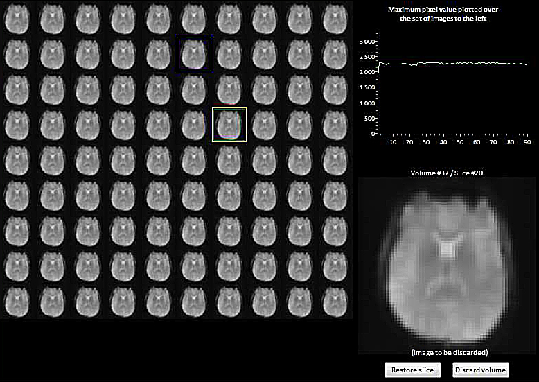
Montage view of source data from an fMRI series
allowing recognition and exclusion of aberrant individual slices
Quality Assurance: Inspection of source images. Individual slices in an fMRI acquisition commonly suffer from random variations in average signal intensity, noise spikes, ghosts, and data glitches. These may result from physiological sources (patient motion, respiration, cardiac pulsations, anxiety, drowsiness, drugs) or from the scanner itself (field inhomogeneities, eddy currents, gradient heating, electronics). If unrecognized and included in the data analysis, these may spoil the entire experiment. A quick visual review of all source images together in montage mode is highly recommended to search for and exclude ("scrub") aberrant slices that appear too bright, too dark, or contain artifacts. More sophisticated graphical and semi-automated methods to identify outlier data are also available. 

epi distortion
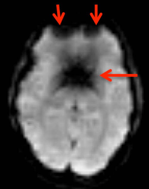
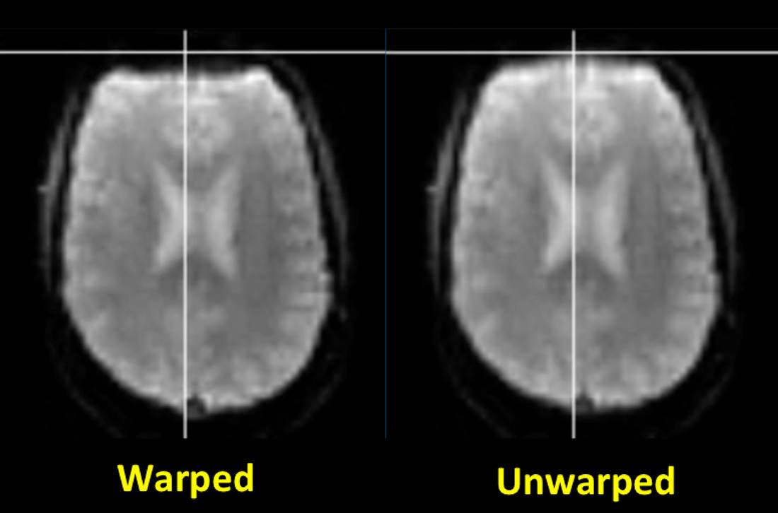
Distortion correction. Common fMRI/BOLD sequences acquire gradient echoes and hence are sensitive to magnetic inhomogeneity (T2*) effects. These cause spatial distortions and signal dropout especially near the skull base, typically affecting the anterior frontal and temporal lobes.
Field mapping and "unwarping" methods (described in the Advanced Discussion) are available to reduce these distortions. Although these techniques may be required for sophisticated neuropsychological experiments, they are not commonly used for basic eloquent cortex mapping in clinical fMRI studies. 
Slice timing correction. Most fMRI studies acquire one slice at a time, meaning that the signal recorded from one slice may be offset in time by up to several seconds when compared to another. The situation is even further complicated depending on whether the slices have been acquired in sequential (1,2,3,4,5,6...) or in interleaved (1,3,5,..2,4,6...) order and whether simultaneous multi-slice imaging has been employed.  Although slice timing differences may not be important for simple block design experiments, they can impart considerable errors in rapid, event-related fMRI studies if not accounted for. 
Slice timing correction fMRI
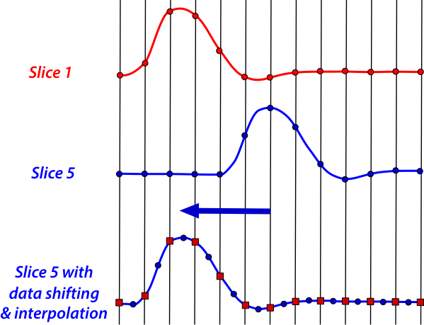
Two basic strategies have been developed for slice timing correction. Data shifting is the most commonly used method, where recorded points are moved to reflect their proper offset from the time of the stimulus. This method requires interpolation of points to fit the fixed, TR-based timing grid and thus produces some blurring and degradation of the data. An alternate (post-processing) strategy is model shifting, where the expected location of the hemodynamic response function (HRF) is varied, treating slice location as an additional independent variable in the subsequent statistical analysis. Sometimes temporal derivatives of the HRF are also incorporated in the model to improve accuracy. 

mean squared error motion correction fmri
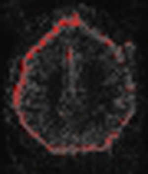
Residual mean-squared error
after motion correction
Motion Correction.  Head motion is the largest source of error in fMRI studies, and a variety of strategies have been developed to cope with this problem. Immobilization of the head using padding and straps is essential; even more rigid restrictions using bite bars and masks are occasionally employed. Proper coaching and training of the subject prior to imaging is important. Prospective motion correction using navigator echoes may be performed but more commonly motion correction is done retrospectively.

motion fmri
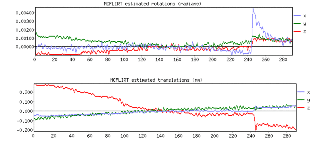
Rotation and translation plots of the 6 parameters in a motion correction algorithm
Note sudden head movement jump at acquisition #245. (Courtesy Chris Rorden)
The standard retrospective motion correction method considers the head as a rigid body with three directions of translation (displacement) and three axes of rotation. A single functional volume of a run is chosen as the reference to which runs in all other volumes are aligned. An iterative procedure is performed in which each volume is rotated and aligned with the reference, with the goal to minimize a cost function (such as the mean-squared difference). This iterative adjustment terminates once no further improvement can be achieved. All major fMRI analysis packages produce line plots allowing visual inspection of how translation and rotation parameters change from volume to volume (see figure above).

fmri temporal filtering
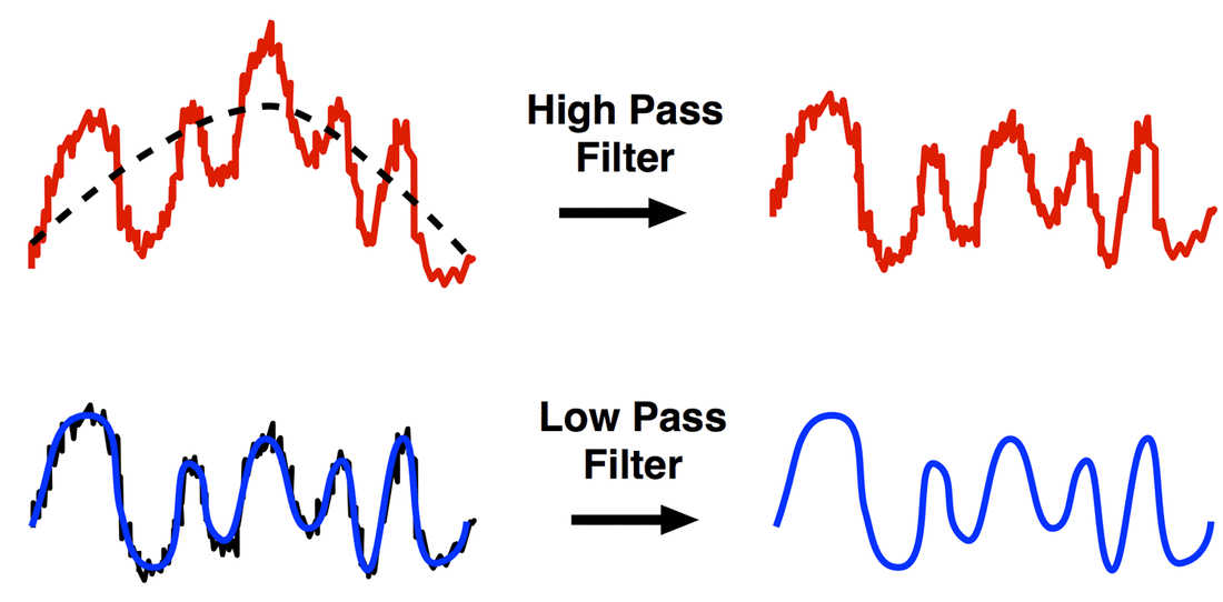
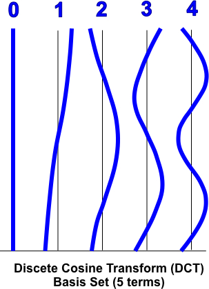
Temporal Filtering. fMRI data nearly always exhibit slow wandering of the baseline signal over time as well as rapid fluctuations due to noise. The removal of low frequency drifts is known as detrending. Detrending may be accomplished using either high-pass filtering after Fourier transformation or by time-domain averaging methods. Alternatively, gradual drifts can be removed later in the data analysis pipeline by adding a set of confound predictors (such as a discrete cosine transform basis set) to account for low-frequency fluctuations. High-frequency signal fluctuations (AKA "noise") can be removed by low-pass filtering. Low-pass filtering is generally not recommended for most studies, however, since it may distort estimation of individual HRFs and reduce the fMRI signals of interest.

spatial smoothing fMRI
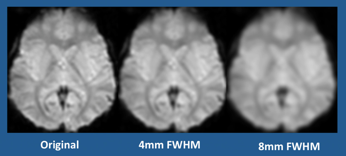
Spatial smoothing using Gaussian filters with various kernel widths
(FWHM = full width half maximum)
Spatial Smoothing.  Spatial smoothing is the averaging of signals from adjacent voxels. This improves the signal-to-noise ratio (SNR) but decreases spatial resolution, blurs the image, and smears activated areas into adjacent voxels. The process can be justified because closely neighboring brain voxels are usually inherently correlated in their function and blood supply. The standard method is to convolve ("multiply") the fMRI data with a 3D Gaussian kernel ("filter") that averages signals from neighboring voxels with weights that decrease with increasing distance from the target voxel. The optimal kernel size is disputed, depending on factors such as slice thickness and in-plane resolution and the need for spatial separation of small activation regions. In practice, the full width half maximum (FWHM) value of the Gaussian spatial filter is typically set to about 4-6 mm for single subject studies and to about 6-8 mm for multi-subject analyses. 

Advanced Discussion (show/hide)»

Additional Comments About fMRI Pre-Processing
Quality Assurance
​Although all slices should be quickly assessed visually, the initial slices of an fMRI acquisition should be especially scrutinized. This is the time when tissue magnetization may not have not yet reached a steady-state equilibrium and signal intensity may appear artificially high. Most scanners perform one or more “dummy scans” and discard the first 3-5 slices, but this is worth checking. For some experiments it may be desirable to censor even more initial slices (e.g., to allow a subject to acclimatize to loud scanner sounds). Graphical methods (illustrated below) permit rapid identification of outlier data. High-signal outliers result in false areas of activation; low-signal outliers create false areas of deactivation. Additional time series diagnostics include mean voxel intensity, variance of voxel intensity, and temporal signal-to-noise per slice.
fMRI data spikes

Above: Plot of mean signal per slice allows quick identification of outlier points

Below: Percent of outlier data points by slice and experiment
fmri data outliers
An option called "Mean Intensity Adjustment" (or one with similar wording) is available in most fMRI software. Its purpose is to remove global fluctuations in average background signal from slice to slice. Before the current era of fully digital RF multi-transmit and receiver chains, this option was more widely used, but is now largely unnecessary for modern scanners operating at 3T or below. Intensity normalization may still be needed for fMRI studies at 7T or higher where RF-inhomogeneities can be more problematic.
Distortion Correction
Field mapping and unwarping. Basic rapid field mapping is commonly performed as a normal part of automated prescan procedures. In the simplest case a low-resolution dual echo sequence with relatively short TE's is first performed, with computation of magnitude and phase-difference images. The phase-difference images require unwrapping as all phase measurements (however large) are "wrapped" (compressed) over the range of 0°−360°. After phase unwrapping field map values are used to calculate corrective pixel shifts in the phase-encode direction, a process known as unwarping (not to be confused with unwrapping above!). Because field map artifacts and noise create problems, some spatial smoothing (often Gaussian) is typically performed as well as masking to exclude voxels outside of the brain. More sophisticated field mapping and unwarping techniques include those that continuously update during data acquisition.
Picture
Field mapping (magnitude images on left, unwrapped
phase images on right). Courtesy of Chris Rorden.
unwarping fMRI
Image unwarping after phase mapping (Modified from figure courtesy of Chris Rorden)
Z-shimming. T2*-dephasing creates inhomogeneity in the z-(slice-select)-direction as well as in plane (xy-direction). To restore the signal a compensating gradient along the z-axis is applied to be sure the k-space trajectory returns to the origin at time TE. As commonly implemented, images are collected using 3-4 different positive and negative z-gradients applied immediately after the RF-pulse and then combined using a square root sum of squares approach. The method may also be applied to reduce in-plane artifacts in the phase-encode direction, and a 3D-implementation has also been developed. A major disadvantage of z-shimming is the time penalty, meaning a longer TR with fewer slices and decreased temporal resolution. Additionally, portions of the original image not suffering from susceptibility-induced signal loss can be dephased by the z-shimming gradients and potentially made worse.
It should be noted that distortion correction techniques such as field mapping, unwarping, and z-shimming cannot restore fMRI signals lost due to dephasing. These techniques merely attempt to reassign recorded signals to the proper points in space from which they arose.
Slice-Timing Correction
Interpolation of the time-shifted data points may be performed by nearest neighbor, tri-linear, multipoint spline, or sinc approximations. Nearest neighbor and linear interpolation are the fastest, but introduce more smoothing artifacts; cubic/multipoint B-spline or (Hanning-windowed) sinc methods are generally preferred. These latter techniques tend to spread the artifact out over a wider range of nearby voxels. Whether to do slice time-correction as a pre-processing step vs post-processing HRF model shifting remains in dispute. Likewise, if the pre-processing option is chosen, whether to do slice-timing correction before or after motion correction is also not agreed upon, and may depend on the expected degree of motion as well as slice order.
Motion Correction
The iterative minimization procedure used for fMRI motion correction schemes is typically a nonlinear least squares routine (e.g., Levenberg-Marquardt). Possible errors in this process may result from finding only a local (rather than global) minimum of the cost function, leading to suboptimal results. Once motion parameters for realignment have been determined, they are applied to create a new 3D motion-corrected data set. Creation of the corrected data set requires spatial interpolation, as the new data points typically fall in between the original uncorrected data points. This process may be computationally-intensive, so usually some combination of (fast) linear interpolation is used during the initial motion correction steps, followed by a more time-consuming interpolation method (such as windowed sinc) for the final spatial transformation.

Typically head motions are relatively small (<2 mm) during normal fMRI experiments, so the assumptions underlying this rigid body approach are justified. However, sudden abrupt head motion (as seen near volume 245 in the figure above) will violate these conditions and may not produce an appropriate correction for motion.

Rigid body transformations cannot compensate for non-linear effects. These include field inhomogeneity effects, motion during slice acquisition, interpolation artifacts, and spin-excitation history effects. Field inhomogeneity effects may be the most important. Even though rigid head motion can be corrected in image space, head displacements affect magnetic field homogeneity and shimming, so even with perfect realignment some motion-related errors persist. These additional sources of residual motion error are often referred to as the residual variance and may be addressed in part by unwarping (described above).
Temporal Filtering
Low frequency drifts in fMRI data are very common, and if not accounted for, will severely reduce the power of the statistical analysis. They will also invalidate event-related averaging, which assumes a stationary level of signal over the course of an experiment. Accordingly, removing these drifts is a mandated step for every fMRI study. This must be done carefully, however, as true condition-related signal changes may be inadvertently removed by this process if improperly applied.

Typically, low frequency drifts are removed as part of the preprocessing pipeline. They may also be removed during the post-processing/statistical analysis phase by incorporating them as “nuisance predictors” in the General Linear Model (GLM). Both approaches are commonly used (although not together) with generally similar results.
​
If the pre-processing route is chosen, then a Fourier frequency filtering method is typically employed. Here, the fMRI signal time course for a voxel is transformed into the frequency domain using a Fast Fourier Transform (FFT). A certain group of low frequencies (say 1-3 cycles) is removed, and the filtered data retransformed back into the time domain.
Because FFT frequency filtering does not work well with purely linear trends, several fMRI software packages first remove linear trends in the time domain with a simple regression tool before transforming into the frequency domain. Additionally Fourier-based filtering techniques may also introduce spurious autocorrelations into the data.

Accounting for low frequency drifts can also be done as a post-processing step, including them as confound predictors in the GLM. Some investigators prefer this approach. In brief, 3-5 additional columns are added to the GLM design matrix (X) composed of low-frequency waves. These may be a low frequency Fourier basis set of sines and cosines plus a constant linear trend term or a slightly more sophisticated (but closely related) discrete cosine transform (DCT) set.
Discrete Cosine Transform Basis Set

References
     Ashburner J, Friston KJ. Rigid body registration. In: Frackowiak RSJ (ed). Human Brain Function, 2nd ed. London: Academic Press, 2004. (pdf made available on-line by the authors at http://www.fil.ion.ucl.ac.uk/spm/doc/books/hbf2/) 
    
Berman H, Mulders J, Goebel R. Anatabacus v1.1: Calculator for image distortions for Brain Voyager QX, 2014. (Detailed manual describing procedures for distortion correction using Brain Voyager, available from this link.  Includes useful information about how to compute field maps and other practical measures for reducing susceptibility distortions.)
     Chen JE, Glover GH. Functional magnetic imaging methods. Neuropsychol Rev 2015; 25:289-313.
     Friston KJ, Josephs O, Zarahn E, et al. To smooth or not to smooth? Bias and efficiency in fMRI time-series analysis. NeuroImage 2000; 12: 196-208. (discussion of temporal smoothing, including bandpass filtering and whitening).
     Maclaren J, Herbst M, Speck O, Zaitsev M. Prospective motion correction in brain imaging: a review. Magn Reson Med 2013; 69: 621–636.
     
Mikl M, Maraček R, Hluštík P, et al. Effects of spatial smoothing on fMRI group inferences. Magn Reson Imaging 2008; 26:490-503.
     Sladky R, Friston KJ, Tröstl J, et al. Slice-timing effects and their correction in functional MRI. Neuroimage 2011; 58:588-594.
     Tanabe J Miller D, Tregellas J, et al. Comparison of detrending methods for optimal fMRI preprocessing. NeuroImage 2002; 15:902–907.
     The FIL Methods Group. SPM12 Manual. Welcome Trust Center for Neuroimaging, London, 2016. (describes preprocessing steps available in the SPM program).

Related Questions
     How is fMRI data processed and analyzed? 

←  Previous Question
Next Question  →
↑ Complete List of Questions ↑
© 2024 AD Elster, ELSTER LLC
All rights reserved.   
MRIquestions.com - Home
Donate
Please help keep this site free for everyone in the world!