Real v Imaginary SignalsWhat is the difference between real and imaginary signals? How can one be more real than another?
|
|
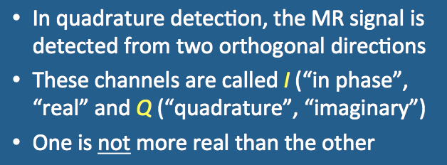
Quadrature detection of the MR signal
|
As described in a previous Q&A, the MR signal is an induced current generated by precession of the net magnetization (M) after stimulation by an RF- pulse. The signal is commonly detected in quadrature using receiver coils sensitive to magnetic flux in two orthogonal directions. Output channels, denoted I and Q (for "in phase" and "quadrature" respectively), send their respective signals along separate digitization and amplifier pathways. These signals are ultimately demodulated, processed, and recombined to create the final MR image.
|
The quadrature receiver coils are measuring the same precessing magnetization (M) from two different perspectives. The signals in the I and Q channels, therefore, should theoretically be identical except for a 90º-phase shift between them. The second coil permits knowledge of the exact position of M and hence its direction of its rotation (i.e., positive vs negative frequency). Another way to think of this is that with quadrature detection you are "listening" to the MR signal in "stereo". The same "music" is coming out of each speaker but you can now tell that the violins are on the left and the horns are on the right.
In reality the signals from the I and Q channels are not phase-shifted exact copies of one another because they also contain noise. Unlike the signals, noise in the two channels is independent and uncorrelated. Thus quadrature detection offers an increase in signal-to-noise by a factor of √2 = 1.41 over detection by a single linear receiver coils.
|
The MR signal can be represented as a vector with real (Re) and imaginary (Im) components recorded from the I and Q channels respectively. An equivalent/alternative representation of the signal is as a complex number
Signal = (Re, Im) = Re + i Im
where i² = −1, the imaginary unit. Its magnitude and phase can be calculated by simple trigonometry.
|
Representation of the MR signal in terms of real and imaginary components.
|
The designation of the I and Q signal channels as "real" and "imaginary" is entirely arbitrary. The signal from one channel is no more or less "real" than that from the other channel.
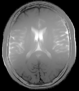
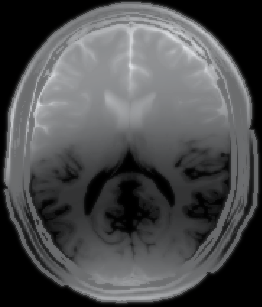
The signal data may be reconstructed in several ways: (1) as a "real" image, (2) as an "imaginary" image, (3) as a magnitude image, or (4) as a phase image. For illustrative purposes I have processed the data from the I and Q channels separately to demonstrate the contribution of each component more concretely. The horizontal bands are phase-shifted from each other by 90° and reflect a baseline phase error across the imaged volume.
Real
|
Imaginary
|
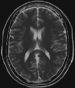
Magnitude
|

Phase
|
In clinical practice, separate I and Q channel images are never obtained, with magnitude images being used nearly exclusively for diagnosis. Phase-images are occasionally generated in clinical MRI for the depiction of flow and characterization of susceptibility-induced distortions.
References
Gudbjartsson H, Patz. The Rician distribution of noisy MRI data. Magn Reson Med 1995; 34:910-914.
Stormont RS, Anas MC, Pelc NJ. Radio frequency receiver for a NMR instrument. US Patent #4992736A, published 12 Feb 1991.
Stormont RS, Anas MC, Pelc NJ. Radio frequency receiver for a NMR instrument. US Patent #4992736A, published 12 Feb 1991.
Related Questions
What is the difference between linearly polarized (LP) and a circularly polarized (CP) coils?
What is the difference between linearly polarized (LP) and a circularly polarized (CP) coils?