Questions and Answers ​in MRI
  • Home
  • Complete List of Questions
  • …Magnets & Scanners
    • Basic Electromagnetism >
      • What causes magnetism?
      • What is a Tesla?
      • Who was Tesla?
      • What is a Gauss?
      • How strong is 3.0T?
      • What is a gradient?
      • Aren't gradients coils?
      • What is susceptibility?
      • How to levitate a frog?
      • What is ferromagnetism?
      • Superparamagnetism?
    • Magnets - Part I >
      • Types of magnets?
      • Brands of scanners?
      • Which way does field point?
      • Which is the north pole?
      • Low v mid v high field?
      • Advantages to low-field?
      • Disadvantages?
      • What is homogeneity?
      • Why homogeneity?
      • Why shimming?
      • Passive shimming?
      • Active shimming?
    • Magnets - Part II >
      • Superconductivity?
      • Perpetual motion?
      • How to ramp?
      • Superconductive design?
      • Room Temp supercon?
      • Liquid helium use?
      • What is a quench?
      • Is field ever turned off?
      • Emergency stop button?
    • Gradients >
      • Gradient coils?
      • How do z-gradients work?
      • X- and Y- gradients?
      • Open scanner gradients?
      • Eddy current problems?
      • Active shielded gradients?
      • Active shield confusion?
      • What is pre-emphasis?
      • Gradient heating?
      • Gradient specifications?
      • Gradient linearity?
    • RF & Coils >
      • Many kinds of coils?
      • Radiofrequency waves?
      • Phase v frequency?
      • RF Coil function(s)?
      • RF-transmit coils?
      • LP vs CP (Quadrature)?
      • Multi-transmit RF?
      • Receive-only coils?
      • Array coils?
      • AIR Coils?
    • Site Planning >
      • MR system layout?
      • What are fringe fields?
      • How to reduce fringe?
      • Magnetic shielding?
      • Need for vibration testing?
      • What's that noise?
      • Why RF Shielding?
      • Wires/tubes thru wall?
  • ...Safety and Screening
    • Overview >
      • ACR Safety Zones?
      • MR safety screening?
      • Incomplete screening?
      • Passive v active implants?
      • Conditional implants?
      • Common safety issues?
      • Projectiles?
      • Metal detectors?
      • Pregnant patients?
      • Postop, ER & ICU patients?
      • Temperature monitoring?
      • Orbital foreign bodies?
      • Bullets and shrapnel?
    • Static Fields >
      • "Dangerous" metals?
      • "Safe" metals?
      • Magnetizing metal?
      • Object shape?
      • Forces on metal?
      • Most dangerous place?
      • Force/torque testing?
      • Static field bioeffects?
      • Dizziness/Vertigo?
      • Flickering lights?
      • Metallic taste?
    • RF Fields >
      • RF safety overview?
      • RF biological effects?
      • What is SAR?
      • SAR limits?
      • Operating modes?
      • How to reduce SAR?
      • RF burns?
      • Estimate implant heating?
      • SED vs SAR?
      • B1+rms vs SAR?
      • Personnel exposure?
      • Cell phones?
    • Gradient Fields >
      • Gradient safety overview
      • Acoustic noise?
      • Nerve stimulation?
      • Gradient vs RF heating?
    • Safety: Neurological >
      • Aneurysm coils/clips?
      • Shunts/drains?
      • Pressure monitors/bolts?
      • Deep brain stimulators?
      • Spinal cord stimulators?
      • Vagal nerve stimulators?
      • Cranial electrodes?
      • Carotid clamps?
      • Peripheral stimulators?
      • Epidural catheters?
    • Safety: Head & Neck >
      • Additional orbit safety?
      • Cochlear Implants?
      • Bone conduction implants?
      • Other ear implants?
      • Dental/facial implants?
      • ET tubes & airways?
    • Safety: Chest & Vascular >
      • Breast tissue expanders?
      • Breast biopsy markers?
      • Airway stents/valves/coils?
      • Respiratory stimulators?
      • Ports/vascular access?
      • Swan-Ganz catheters?
      • IVC filters?
      • Implanted infusion pumps?
      • Insulin pumps & CGMs?
      • Vascular stents/grafts?
      • Sternal wires/implants?
    • Safety: Cardiac >
      • Pacemaker dangers?
      • Pacemaker terminology?
      • New/'Safe" Pacemakers?
      • Old/Legacy Pacemakers?
      • Violating the conditions?
      • Epicardial pacers/leads?
      • Cardiac monitors?
      • Heart valves?
      • Miscellaneous CV devices?
    • Safety: Abdominal >
      • PIllCam and capsules?
      • Gastric pacemakers?
      • Other GI devices?
      • Contraceptive devices?
      • Foley catheters?
      • Incontinence devices?
      • Penile Implants?
      • Sacral nerve stimulators?
      • GU stents and other?
    • Safety: Orthopedic >
      • Orthopedic hardware?
      • External fixators?
      • Traction and halos?
      • Bone stimulators?
      • Magnetic rods?
  • …The NMR Phenomenon
    • Spin >
      • What is spin?
      • Why I = ½, 1, etc?
      • Proton = nucleus = spin?
      • Predict nuclear spin (I)?
      • Magnetic dipole moment?
      • Gyromagnetic ratio (γ)?
      • "Spin" vs "Spin state"?
      • Energy splitting?
      • Fall to lowest state?
      • Quantum "reality"?
    • Precession >
      • Why precession?
      • Who was Larmor?
      • Energy for precession?
      • Chemical shift?
      • Net magnetization (M)?
      • Does M instantly appear?
      • Does M also precess?
      • Does precession = NMR?
    • Resonance >
      • MR vs MRI vs NMR?
      • Who discovered NMR?
      • How does B1 tip M?
      • Why at Larmor frequency?
      • What is flip angle?
      • Spins precess after 180°?
      • Phase coherence?
      • Release of RF energy?
      • Rotating frame?
      • Off-resonance?
      • Adiabatic excitation?
      • Adiabatic pulses?
    • Relaxation - Physics >
      • Bloch equations?
      • What is T1?
      • What is T2?
      • Relaxation rate vs time?
      • Why is T1 > T2?
      • T2 vs T2*?
      • Causes of Relaxation?
      • Dipole-dipole interactions?
      • Chemical Exchange?
      • Spin-Spin interactions?
      • Macromolecule effects?
      • Which H's produce signal?
      • "Invisible" protons?
      • Magnetization Transfer?
      • Bo effect on T1 & T2?
      • How to predict T1 & T2?
    • Relaxation - Clincial >
      • T1 bright? - fat
      • T1 bright? - other oils
      • T1 bright? - cholesterol
      • T1 bright? - calcifications
      • T1 bright? - meconium
      • T1 bright? - melanin
      • T1 bright? - protein/mucin
      • T1 bright? - myelin
      • Magic angle?
      • MT Imaging/Contrast?
  • …Pulse Sequences
    • MR Signals >
      • Origin of MR signal?
      • Free Induction Decay?
      • Gradient echo?
      • TR and TE?
      • Spin echo?
      • 90°-90° Hahn Echo?
      • Stimulated echoes?
      • STEs for imaging?
      • 4 or more RF-pulses?
      • Partial flip angles?
      • How is signal higher?
      • Optimal flip angle?
    • Spin Echo >
      • SE vs Multi-SE vs FSE?
      • Image contrast: TR/TE?
      • Opposite effects ↑T1 ↑T2?
      • Meaning of weighting?
      • Does SE correct for T2?
      • Effect of 180° on Mz?
      • Direction of 180° pulse?
    • Inversion Recovery >
      • What is IR?
      • Why use IR?
      • Phase-sensitive IR?
      • Why not PSIR always?
      • Choice of IR parameters?
      • TI to null a tissue?
      • STIR?
      • T1-FLAIR
      • T2-FLAIR?
      • IR-prepped sequences?
      • Double IR?
    • Gradient Echo >
      • GRE vs SE?
      • Multi-echo GRE?
      • Types of GRE sequences?
      • Commercial Acronyms?
      • Spoiling - what and how?
      • Spoiled-GRE parameters?
      • Spoiled for T1W only?
      • What is SSFP?
      • GRASS/FISP: how?
      • GRASS/FISP: parameters?
      • GRASS vs MPGR?
      • PSIF vs FISP?
      • True FISP/FIESTA?
      • FIESTA v FIESTA-C?
      • DESS?
      • MERGE/MEDIC?
      • GRASE?
      • MP-RAGE v MR2RAGE?
    • Susceptibility Imaging >
      • What is susceptibility (χ)?
      • What's wrong with GRE?
      • Making an SW image?
      • Phase of blood v Ca++?
      • Quantitative susceptibility?
    • Diffusion: Basic >
      • What is diffusion?
      • Iso-/Anisotropic diffusion?
      • "Apparent" diffusion?
      • Making a DW image?
      • What is the b-value?
      • b0 vs b50?
      • Trace vs ADC map?
      • Light/dark reversal?
      • T2 "shine through"?
      • Exponential ADC?
      • T2 "black-out"?
      • DWI bright causes?
    • Diffusion: Advanced >
      • Diffusion Tensor?
      • DTI (tensor imaging)?
      • Whole body DWI?
      • Readout-segmented DWI?
      • Small FOV DWI?
      • IVIM?
      • Diffusion Kurtosis?
    • Fat-Water Imaging >
      • Fat & Water properties?
      • F-W chemical shift?
      • In-phase/out-of-phase?
      • Best method?
      • Dixon method?
      • "Fat-sat" pulses?
      • Water excitation?
      • STIR?
      • SPIR?
      • SPAIR v SPIR?
      • SPIR/SPAIR v STIR?
  • …Making an Image
    • From Signals to Images >
      • Phase v frequency?
      • Angular frequency (ω)?
      • Signal squiggles?
      • Real v Imaginary?
      • Fourier Transform (FT)?
      • What are 2D- & 3D-FTs?
      • Who invented MRI?
      • How to locate signals?
    • Frequency Encoding >
      • Frequency encoding?
      • Receiver bandwidth?
      • Narrow bandwidth?
      • Slice-selective excitation?
      • SS gradient lobes?
      • Cross-talk?
      • Frequency encode all?
      • Mixing of slices?
      • Two slices at once?
      • Simultaneous Multi-Slice?
    • Phase Encoding >
      • Phase-encoding gradient?
      • Single PE step?
      • What is phase-encoding?
      • PE and FE together?
      • 2DFT reconstruction?
      • Choosing PE/FE direction?
    • Performing an MR Scan >
      • What are the steps?
      • Automatic prescan?
      • Routine shimming?
      • Coil tuning/matching?
      • Center frequency?
      • Transmitter gain?
      • Receiver gain?
      • Dummy cycles?
      • Where's my data?
      • MR Tech qualifications?
    • Image Quality Control >
      • Who regulates MRI?
      • Who accredits?
      • Mandatory accreditation?
      • Routine quality control?
      • MR phantoms?
      • Geometric accuracy?
      • Image uniformity?
      • Slice parameters?
      • Image resolution?
      • Signal-to-noise?
      • Ghosting?
  • …K-space & Rapid Imaging
    • K-space (Basic) >
      • What is k-space?
      • Parts of k-space?
      • What does "k" stand for?
      • Spatial frequencies?
      • Locations in k-space?
      • Data for k-space?
      • Why signal ↔ k-space?
      • Spin-warp imaging?
      • Big spot in middle?
      • K-space trajectories?
      • Radial sampling?
    • K-space (Advanced) >
      • K-space grid?
      • Negative frequencies?
      • Field-of-view (FOV)
      • Rectangular FOV?
      • Partial Fourier?
      • Phase symmetry?
      • Read symmetry?
      • Why not use both?
      • ZIP?
    • Rapid Imaging (FSE &EPI) >
      • What is FSE/TSE?
      • FSE parameters?
      • Bright Fat?
      • Other FSE differences?
      • Dual-echo FSE?
      • Driven equilibrium?
      • Reduced flip angle FSE?
      • Hyperechoes?
      • SPACE/CUBE/VISTA?
      • Echo-planar imaging?
      • HASTE/SS-FSE?
    • Parallel Imaging (PI) >
      • What is PI?
      • How is PI different?
      • PI coils and sequences?
      • Why and when to use?
      • Two types of PI?
      • SENSE/ASSET?
      • GRAPPA/ARC?
      • CAIPIRINHA?
      • Compressed sensing?
      • Noise in PI?
      • Artifacts in PI?
  • …Contrast Agents
    • Contrast Agents: Physics >
      • Why Gadolinium?
      • Paramagnetic relaxation?
      • What is relaxivity?
      • Why does Gd shorten T1?
      • Does Gd affect T2?
      • Gd & field strength?
      • Best T1-pulse sequence?
      • Triple dose and MT?
      • Dynamic CE imaging?
      • Gadolinium on CT?
    • Contrast Agents: Clinical >
      • So many Gd agents!
      • Important properties?
      • Ionic v non-ionic?
      • Intra-articular/thecal Gd?
      • Gd liver agents (Eovist)?
      • Mn agents (Teslascan)?
      • Feridex & Liver Agents?
      • Lymph node agents?
      • Ferumoxytol?
      • Blood pool (Ablavar)?
      • Bowel contrast agents?
    • Contrast Agents: Safety >
      • Gadolinium safety?
      • Allergic reactions?
      • Renal toxicity?
      • What is NSF?
      • NSF by agent?
      • Informed consent for Gd?
      • Gd protocol?
      • Is Gd safe in infants?
      • Reduced dose in infants?
      • Gd in breast milk?
      • Gd in pregnancy?
      • Gd accumulation?
      • Gd deposition disease?
  • …Cardiovascular and MRA
    • Flow effects in MRI >
      • Defining flow?
      • Expected velocities?
      • Laminar v turbulent?
      • Predicting MR of flow?
      • Time-of-flight effects?
      • Spin phase effects?
      • Flow void?
      • Why GRE ↑ flow signal?
      • Slow flow v thrombus?
      • Even-echo rephasing?
      • Flow-compensation?
      • Flow misregistration?
    • MR Angiography - I >
      • MRA methods?
      • Dark vs bright blood?
      • Time-of-Flight (TOF) MRA?
      • 2D vs 3D MRA?
      • MRA parameters?
      • Magnetization Transfer?
      • Ramped flip angle?
      • MOTSA?
      • Fat-suppressed MRA?
      • TOF MRA Artifacts?
      • Phase-contrast MRA?
      • What is VENC?
      • Measuring flow?
      • 4D Flow Imaging?
      • How accurate?
    • MR Angiography - II >
      • Gated 3D FSE MRA?
      • 3D FSE MRA parameters?
      • SSFP MRA?
      • Inflow-enhanced SSFP?
      • MRA with ASL?
      • Other MRA methods?
      • Contrast-enhanced MRA?
      • Timing the bolus?
      • View ordering in MRA?
      • Bolus chasing?
      • TRICKS or TWIST?
      • CE-MRA artifacts?
    • Cardiac I - Intro/Anatomy >
      • Cardiac protocols?
      • Patient prep?
      • EKG problems?
      • Magnet changes EKG?
      • Gating v triggering?
      • Gating parameters?
      • Heart navigators?
      • Dark blood/Double IR?
      • Why not single IR?
      • Triple IR?
      • Polar plots?
      • Coronary artery MRA?
    • Cardiac II - Function >
      • Beating heart movies?
      • Cine parameters?
      • Real-time cine?
      • Ventricular function?
      • Tagging/SPAMM?
      • Perfusion: why and how?
      • 1st pass perfusion?
      • Quantifying perfusion?
      • Dark rim artifact
    • Cardiac III - Viability >
      • Gd enhancement?
      • TI to null myocardium?
      • PS (phase-sensitive) IR?
      • Wideband LGE?
      • T1 mapping?
      • Iron/T2*-mapping?
      • Edema/T2-mapping?
      • Why/how stress test?
      • Stess drugs/agents?
      • Stress consent form?
  • …MR Artifacts
    • Tissue-related artifacts >
      • Chemical shift artifact?
      • Chemical shift in phase?
      • Reducing chemical shift?
      • Chemical Shift 2nd Kind?
      • In-phase/out-of phase?
      • IR bounce point?
      • Susceptibility artifact?
      • Metal suppression?
      • Dielectric effect?
      • Dielectric Pads?
    • Motion-related artifacts >
      • Why discrete ghosts?
      • Motion artifact direction?
      • Reducing motion artifacts?
      • Saturation pulses?
      • Gating methods?
      • Respiratory comp?
      • Navigator echoes?
      • PROPELLER/BLADE?
    • Technique-related artifacts >
      • Partial volume effects?
      • Slice overlap?
      • Aliasing?
      • Wrap-around artifact?
      • Eliminate wrap-around?
      • Phase oversampling?
      • Frequency wrap-around?
      • Spiral/radial artifacts?
      • Gibbs artifact?
      • Nyquist (N/2) ghosts?
      • Zipper artifact?
      • Data artifacts?
      • Surface coil flare?
      • MRA Artifacts (TOF)?
      • MRA artifacts (CE)?
  • …Functional Imaging
    • Perfusion I: Intro & DSC >
      • Measuring perfusion?
      • Meaning of CBF, MTT etc?
      • DSC v DCE v ASL?
      • How to perform DSC?
      • Bolus Gd effect?
      • T1 effects on DSC?
      • DSC recirculation?
      • DSC curve analysis?
      • DSC signal v [Gd]
      • Arterial input (AIF)?
      • Quantitative DSC?
    • Perfusion II: DCE >
      • What is DCE?
      • How is DCE performed?
      • How is DCE analyzed?
      • Breast DCE?
      • DCE signal v [Gd]
      • DCE tissue parmeters?
      • Parameters to images?
      • K-trans = permeability?
      • Utility of DCE?
    • Perfusion III: ASL >
      • What is ASL?
      • ASL methods overview?
      • CASL?
      • PASL?
      • pCASL?
      • ASL parameters?
      • ASL artifacts?
      • Gadolinium and ASL?
      • Vascular color maps?
      • Quantifying flow?
    • Functional MRI/BOLD - I >
      • Who invented fMRI?
      • How does fMRI work?
      • BOLD contrast?
      • Why does BOLD ↑ signal?
      • Does BOLD=brain activity?
      • BOLD pulse sequences?
      • fMRI Paradigm design?
      • Why "on-off" comparison?
      • Motor paradigms?
      • Visual?
      • Language?
    • Functional MRI/BOLD - II >
      • Process/analyze fMRI?
      • Best fMRI software?
      • Data pre-processing?
      • Registration/normalization?
      • fMRI statistical analysis?
      • General Linear Model?
      • Activation "blobs"?
      • False activation?
      • Resting state fMRI?
      • Analyze RS-fMRI?
      • Network/Graphs?
      • fMRI at 7T?
      • Mind reading/Lie detector?
      • fMRI critique?
  • …MR Spectroscopy
    • MRS I - Basics >
      • MRI vs MRS?
      • Spectra vs images?
      • Chemical shift (δ)?
      • Measuring δ?
      • Backward δ scale?
      • Predicting δ?
      • Size/shapes of peaks?
      • Splitting of peaks?
      • Localization methods?
      • Single v multi-voxel?
      • PRESS?
      • STEAM?
      • ISIS?
      • CSI?
    • MRS II - Clinical ¹H MRS >
      • How-to: brain MRS?
      • Water suppression?
      • Fat suppression?
      • Normal brain spectra?
      • Choice of TR/TE/etc?
      • Hunter's angle?
      • Lactate inversion?
      • Metabolite mapping?
      • Metabolite quantitation?
      • Breast MRS?
      • Gd effect on MRS?
      • How-to: prostate MRS?
      • Prostate spectra?
      • Muscle ¹H-MRS?
      • Liver ¹H-MRS?
      • MRS artifacts?
    • MRS III - Multi-nuclear >
      • Other nuclei?
      • Why phosphorus?
      • How-to: ³¹P MRS
      • Normal ³¹P spectra?
      • Organ differences?
      • ³¹P measurements?
      • Decoupling?
      • NOE?
      • Carbon MRS?
      • Sodium imaging?
      • Xenon imaging?
  • ...Artificial Intelligence
    • AI Part I: Basics >
      • Artificial Intelligence (AI)?
      • What is a neural network?
      • Machine Learning (ML)?
      • Shallow v Deep ML?
      • Shallow networks?
      • Deep network types?
      • Data prep and fitting?
      • Back-Propagation?
      • DL 'Playground'?
    • AI Part 2: Advanced >
      • What is convolution?
      • Convolutional Network?
      • Softmax?
      • Upsampling?
      • Limitations/Problems of AI?
      • Is the Singularity near?
    • AI Part 3: Image processing >
      • AI in clinical MRI?
      • Super-resolution?
  • ...Tissue Properties Imaging
    • MRI of Hemorrhage >
      • Hematoma overview?
      • Types of Hemoglobin?
      • Hyperacute/Oxy-Hb?
      • Acute/Deoxy-Hb?
      • Subacute/Met-Hb?
      • Deoxy-Hb v Met-Hb?
      • Extracellular met-Hb?
      • Chronic hematomas?
      • Hemichromes?
      • Ferritin/Hemosiderin?
      • Subarachnoid blood?
      • Blood at lower fields?
    • T2 cartilage mapping
    • MR Elastography?
    • Synthetic MRI?
    • Amide Proton Transfer?
    • MR thermography?
    • Electric Properties Imaging?
  • Copyright/Legal
    • Copyright Issues
    • Legal Disclaimers
  • Forums/Blogs/Links
  • What's New
  • Self-test Quizzes - NEW!
    • Magnets & Scanners Quiz
    • Safety & Screening Quiz
    • NMR Phenomenon Quiz
    • Pulse Sequences Quiz
    • Making an Image Quiz
    • K-space & Rapid Quiz
    • Contrast & Blood Quiz
    • Cardiovascular & MRA Quiz

Pacemaker Types & Terminology

I'm not a cardiologist. Can you explain some of the pacemaker types and terminology relevant to us working in MRI? 
pacemaker types and terminology
Read MR Safety Disclaimer Before Proceeding
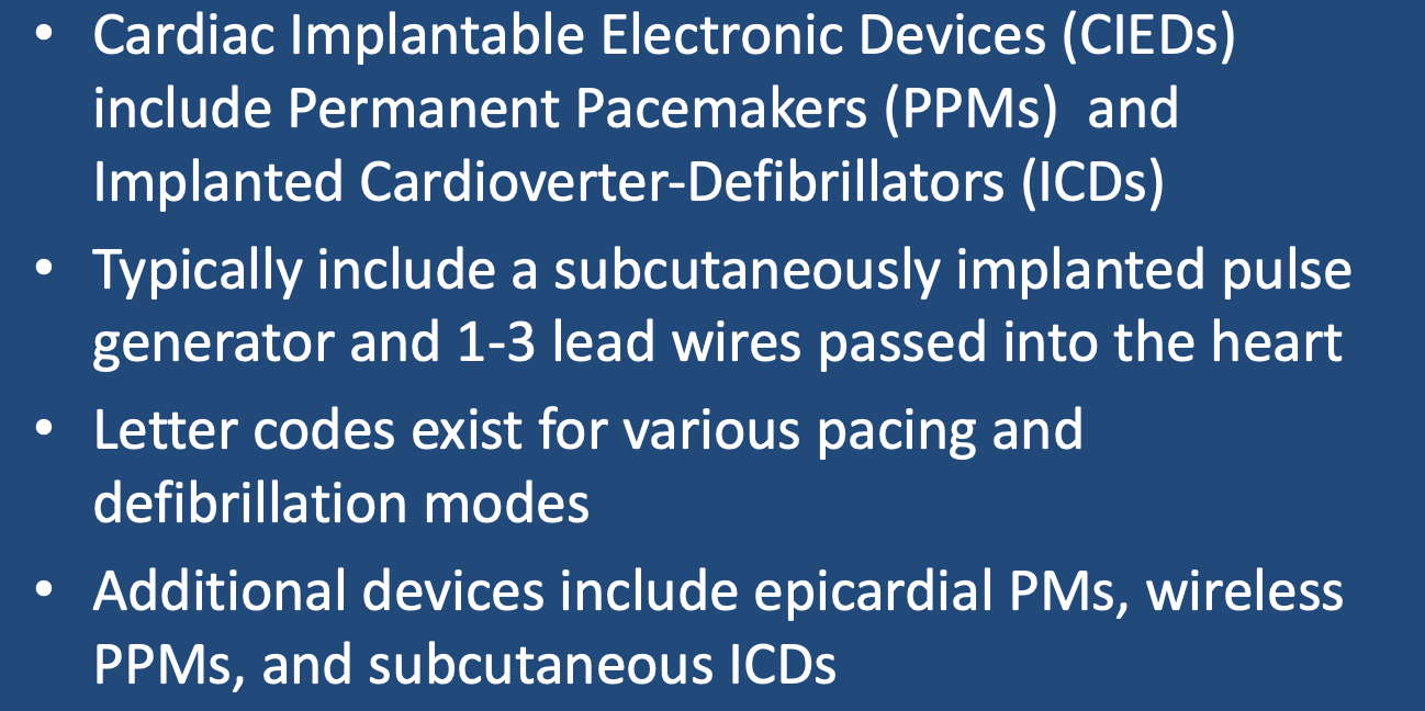
Cardiac Implantable Electronic Devices (CIEDs) are designed to detect and treat disturbances in cardiac rhythm (arrhythmias). CIEDs include both Permanent Pacemakers (PPMs) and Implanted Cardioverter-Defibrillators (ICDs). PPMs deliver relatively mild shocks to the cardiac conduction system and are used primarily for treatment of slow heart rates (bradycardias). ICDs deliver high energy shocks for treatment of rapid heart rates (tachycardias).
Standard CIED Configurations
With the exception of a few uncommon "leadless" and epicardial devices described below, virtually all CIEDs contain the following elements:
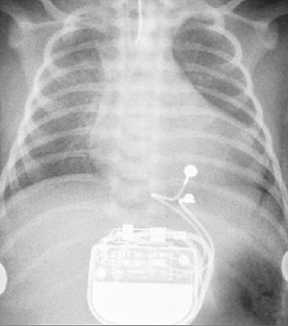
  • A pulse generator (PG), including a lithium-ion battery and electronics housed in a titanium "can", surgically implanted in the chest wall below the clavicle.
  • 1-3 insulated wires leads passed fluoroscopically from the PG to the right heart via the subclavian vein and superior vena cava. Terminal electrodes are affixed to the myocardium by spiral wires or tines. Most modern terminal electrodes are bipolar, meaning they have two bare contact points near their tips, allowing focused sensing of the heart's local electrical activity and accurate delivery of pacing shocks as needed.
Single lead CIEDs typically terminate in the right atrial appendage or right ventricle. They are uncommon and primarily used for bradycardia due to sinus node dysfunction. The overwhelming majority of CIEDs contain two leads (one in the right atrium and one in the right ventricle) for dual-chamber sensing and pacing that follows the natural contraction sequence of the heart. Three-lead CIEDs are primarily used in patients with heart failure or left bundle branch block who require biventricular pacing. The third wire passes through the coronary sinus and into a coronary vein to simulate the left ventricle transvenously. This configuration is commonly referred to as Cardiac Resynchronization Therapy (CRT). Rarely additional leads are present for bundle of His or multisite pacing.
pacemaker wire locations
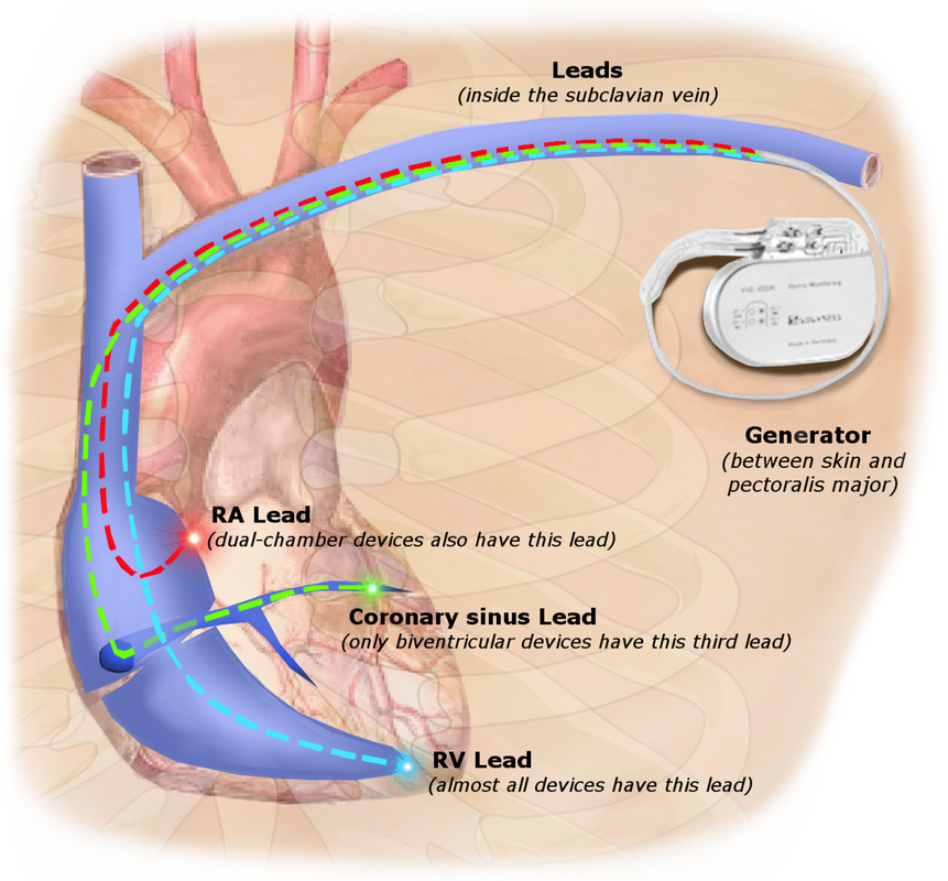
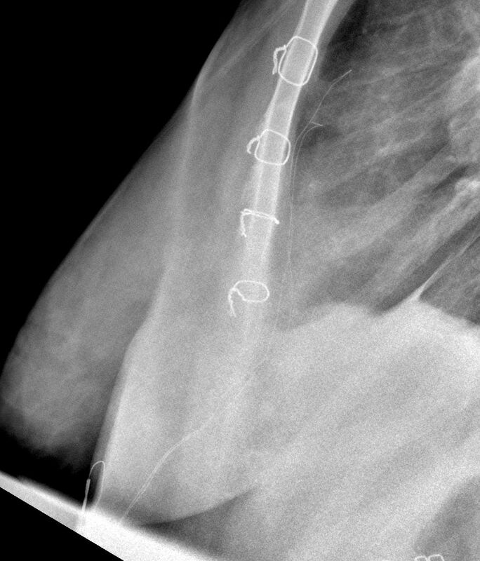
Typical locations for 1-, 2-, and 3-lead pacemakers.
biventricular pacemaker
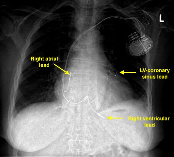
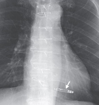
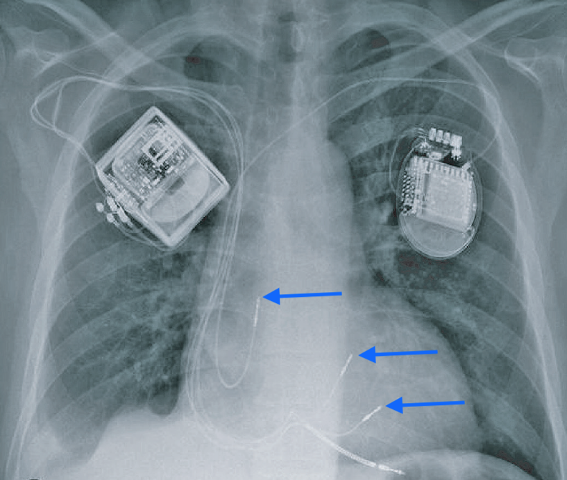
Chest x-ray showing a biventricular PPM with 3 leads
current flows ICD
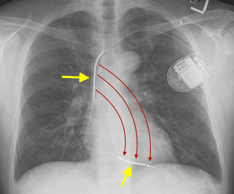
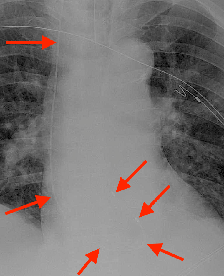
Strong depolarization currents (red arrows) flow between shock coils (yellow arrows) in superior vena cava and right ventricle
CIEDs that operate as high-energy ICDs demonstrate several differences compared to simple PMs. Their PGs are slightly larger, containing bigger batteries and capacitors for storing charge.  They have large surface area electrodes known as shock coils located coaxially along the surface of the pace/sense leads. These shock coils are typically located in the right ventricle and also often in the superior vena cava. They can be recognized on x-ray as focal areas of wire thickening. Strong currents pass between the shock coils to depolarize the heart. Sometimes the housing of the PG functions as an additional current source, the so-called "active can" configuration.  
Several terms are frequently encountered when discussing pacemaker function with regard to MRI:
  • Asynchronous (fixed) pacing: A mode of operation where the PM delivers stimuli at a constant rate regardless of intrinsic cardiac signals.
  • Synchronous (demand) pacing: A mode of operation where the PM delivers a stimulus after a certain time period if no expected cardiac signal is sensed. 
  • Pacing capture threshold: The minimum electrical stimulus measured in millivolts (mV) to consistently depolarize ("capture") the myocardium. This can increase over time as fibrosis builds at the electrode tip.
  • Lead impedance: A measure of resistance to electrical flow within a lead measured in ohms (Ω). A break in the lead can dramatically increase impedance, while a defect in the insulation can result in a short with low impedance.
  • Reed switch: an electrical component within older pulse generators that controls pacing mode and may open/close unpredictably in MRI. 
NBG Codes for Pacemakers​
The North American Society of Pacing and Electrophysiology and British Pacing and Electrophysiology
Groups (NBG) have jointly developed a coding system to describe various pacing modes. The full (revised) code contains 5 letters, but often only the first three are used.
  • The first letter indicates the chamber(s) paced: A=atrium, V=ventricle, or D=dual (both A&V)
  • The second letter indicates in which chamber(s) electrical activity is sensed: A, V, or D. The letter O indicates no sensing is used.
  • The third letter refers to how the PM responds to a sensed cardiac electric signal: T=triggered to give a shock, I= inhibited, D=dual response (any spontaneous A and V activity will inhibit both A&V pacing, but lone atrial activity will trigger a paced ventricular response), and O= no sensing.
  • The fourth and fifth letters are used to denote rate modulation and multisite pacing and are only occasionally used.
Common operational modes include DDD (Atrial and ventricular sequential pacing, with pacing impulses triggered only when programmed intervals are exceeded), VVI (pure ventricular pacing whenever R wave is not detected), DOO (dual asynchronous pacing at constant rate regardless of intrinsic cardiac activity, and OOO (pacemaker disabled).
NBD Codes for Defibrillators
A shortened code by the same societies has been promulgated to reflect the common modes of ICD operation:
  • ICD-S: An ICD with shock capability only
  • ICD-B: An ICD with bradycardia pacing as well as shock
  • ICD-T: An ICD with both tachycardia (and bradycardia) pacing as well as shock
Other Pacing Methods and Devices
  • Temporary Transvenous Pacing is used primarily in emergency situations like acute profound bradycardia. A pacemaker wire is passed through the femoral or internal jugular vein into the heart, while the pulse generator remains outside the body.  Due to the risk of significant lead heating demonstrated in animal models, these devices are considered MR Unsafe.
  • ​Temporary Epicardial Pacing leads are placed in contact with the outer wall of the right heart at the end of open-heart surgery. The wires then extend percutaneously through the chest wall where they are hooked to a small battery-powered pulse generator. The wires are usually removed within a few days to weeks after the patient's postoperative rhythm disturbances have subsided.
  • Permanent Epicardial Pacemakers are used occasionally in pediatric patients with complex congenital heart disease. The pulse generator is typically placed subcutaneously in the upper abdominal wall. 
Picture
"MR Unsafe" temporary transvenous pacemaker inserted via right internal jugular approach
Picture
Temporary epicardial pacing wires
Case courtesy of Dr Jeremy Jones, Radiopaedia.org.
permanent epicardial pacemaker
Permanent epicardial pacemaker in a child with congenital heart disease
From Costa et al under CC-BY
  • Leadless RV Pacemakers are small, self-contained devices implanted fluoroscopically into the right ventricular endocardium, exemplified by the MR Conditional Nanostim™ (Abbott) and Micra™ (Medtronic).  Internal accelerometers allow them to partially sync with right atrial contraction, but essentially they only sense and pace the RV. Because of their small size, they are unable to deliver high-energy shocks for tachyarrythmias. 
Picture
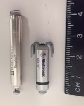
Nanostim™ and Micra™ leadless pacemakers. (From Bhatia et al under CC-BY)
Nanostim® leadless pacemaker
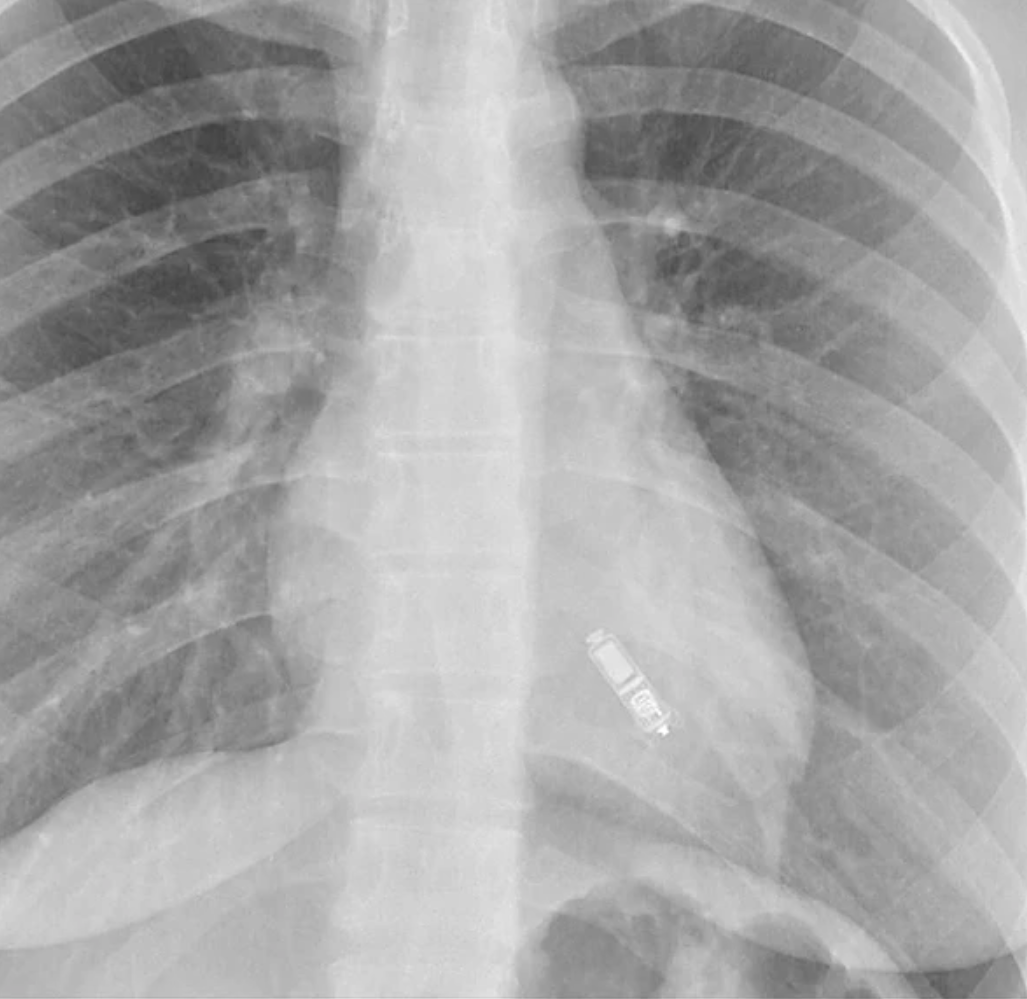
"MR Conditional" Nanostim™ leadless pacemaker
Picture
"MR Conditional" Micra™ leadless pacemaker
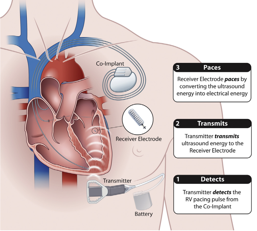
  • Leadless LV Pacemakers.  Only one such device, the MR Conditional WiSE-CRT System (EBR Systems), is commercially available (Europe only).  A tiny (3 x 9 mm) passive electrode-transponder is fluroscopically implanted in the left ventricular endocardium. It converts ultrasound pulses received from a separate subcutaneous generator into electrical stimuli, thus triggering the ventricle. The entire system is not really leadless, since a conventional (wired) dual chamber pacemaker is required to control the right side of the heart.
Wise CRT system diagram
Wise CRT system
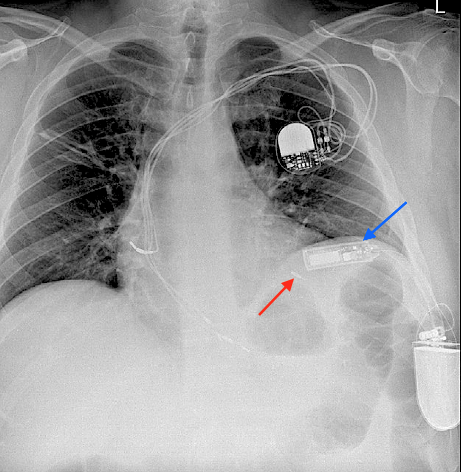
"MR Conditional" WiSE-CRT System showing receiver electrode (red arrow) and ultrasonic transmitter (blue arrow) with subcutaneous pulse generator. Also note standard wired right sided PM.(Both images from Sieniewicz et al under CC-BY)
  • Cardiac Contractility Modulators (CCM). At present there is only one commercial product in this category, the Optimizer™ (Impulse Dynamics), used for patients with refractory heart failure. Resembling a conventional pacemaker, the device is subcutaneously implanted in the subclavicular region with two wires to stimulate the right ventricle. Pulses from Optmizer™ given in four daily 1-hour sessions "exercise" the heart, presumably leading to improved cardiac function. This MR Conditional device requires recharging weekly by an external induction coil.
Picture
"MR Conditional" Optimizer™ CCM device implanted in the right subclavian region with 1 atrial and 2 ventricular electrodes (arrows). Note conventional ICD in left chest.
subcutaneous S-ICD
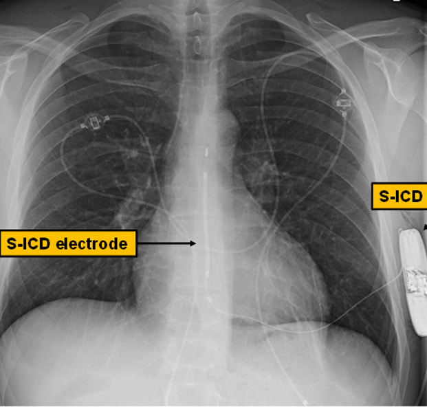
"MR conditional" S-ICD. Vertically oriented electrode is implanted subcutaneously superficial to sternum. (Modified from Tardu under CC-BY)
  • ​Subcutaneous ICDs have shock coils implanted in the anterior chest wall rather than in or on the heart, with the pulse generator can serving as the anode.  The main device in this category is the MR Conditional Emblem™ S-ICD (Boston Scientific). S-ICDs may be devices of choice for young patients or those at risk for intravascular thrombosis.
MRI safety issues concerning conventional CIEDs as well as these other devices are the subject of the next several Q&As.

Advanced Discussion (show/hide)»

No supplementary material yet. Check back soon!

References
     Bhatia N, El-Chami M. Leadless pacemakers: a contemporary review. J Geriatric Cardiol 2018; 15:249-253.  [DIRECT LINK]
     Bernstein A, Camm A, Fisher J, et al. North American Society of Pacing and Electrophysiology policy statement. The NASPE/BPEG defibrillator code. Pacing Clin Electrophysiol 1993; 16:1776–80.     
    Bernstein AD, Daubert J-C, Fletcher RD, et al. The revised NASPE/BPEG generic code for antibradycardia, adaptive-rate, and multisite pacing.  Pacing Clin Electrophysiol 2002; 25:260-264.
     Costa R, da Silva K, Filho MM, Carrillo R. Minimally invasive epicardial pacemaker implantation in neonates with congenital heart block. Arq Bras Cardiol 2017; 109:331-339.
     Mulpuru SK, Madhavan M, McLeod CJ, et al. Cardiac pacemakers: function, troubleshooting, and management.  J Am Coll Cardiol 2017; 69:189-210.  [DOI LINK]
     Mulpuru SK, Madhavan M, McLeod CJ, et al. Advances and future directions in cardiac pacemakers. J Am Coll Cardiol 2017; 69:211–35.  [DOI LINK]
     Sieniewicz BJ, Betts TR, James S, et al. Real-world experience of leadless left ventricular endocardial cardiac resynchronization therapy: A multicenter international registry of the WiSE-CRT pacing system. Heart Rhythm 2020; 17:1291-7.  [DOI LINK]
     Sperzel J, Burri H, Gras D, et al. State of the art of leadless pacing. Europace 2015; 17:1508-1513.

Related Questions
     Do pacemakers stop working in an MRI scanner? Why are older pacemakers dangerous to scan?

←  Previous Question
Next Question  →
↑ Complete List of Questions ↑
© 2024 AD Elster, ELSTER LLC
All rights reserved.   
MRIquestions.com - Home
Donate
Please help keep this site free for everyone in the world!