Invention of functional MRI
Who invented functional MR imaging (fMRI)?
|
|
The term functional MRI (fMRI) usually refers to the imaging of brain activation using various MR techniques. By the late 1980's it was known that regional cerebral blood flow increased near areas of neuronal activity, and low resolution PET studies had already documented this phenomenon in humans. But it remained for a Harvard graduate student, the late John (Jack) Belliveau, working at the Martinos Center at Massachusetts General Hospital, to show that cerebral activation could be imaged using high-resolution MRI.
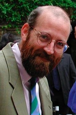
John (Jack) Belliveau
1959-2014 |
The method used by Belliveau et al is now called dynamic susceptibility contrast (DSC) MRI. Echo-planar images of the primary visual cortex were obtained before and after administration of a gadolinium bolus while volunteers viewed a flashing checkerboard pattern through goggles. By subtracting calculated cerebral blood volumes in the stimulated vs non-stimulated states, functional maps were obtained showing increased activity.
|
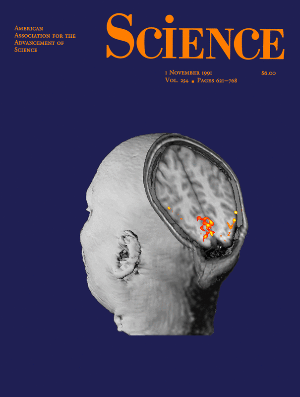
Cover of November 1991 Science with an artist's rendition of the landmark paper by Belliveau et al demonstrating activation of the visual cortex using MRI.
|
This groundbreaking paper in Science excited immediate interest in the neuroscience community, and soon research institutions around the world were clamoring for their own MRI machines. But in 1992, just on the heels of the Belliveau team's discovery, came the demonstration of what would prove to be a superior method for performing fMRI. This technique, known as BOLD (Blood Oxygen Level Dependent) imaging, did not require the use of gadolinium contrast. Instead it exploited different magnetic properties of oxygenated and deoxygenated blood to detect changes in regional blood flow
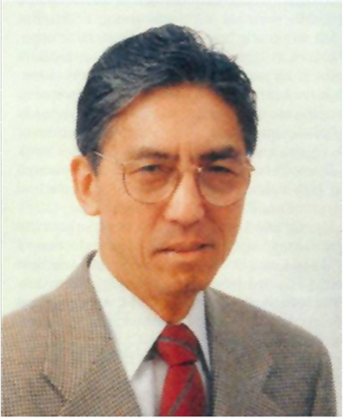
Seiji Ogawa, pioneer of BOLD fMRI, c. 1990
|
The BOLD method was conceived and developed by AT&T Bell Laboratories Scientist Seiji Ogawa, working in conjunction with MRI investigators at the University of Minnesota. BOLD contrast was first demonstrated in rats at 7T in 1990, and was first used for fMRI activation studies in the human visual cortex at 4T during on-off photic stimulation in 1992, published in the Proceedings of the National Academy of Sciences USA. Because it was totally noninvasive (not requiring injection of exogenous contrast material), BOLD allowed multiple cerebral activation experiments to be performed in a single sitting. The BOLD method rapidly supplanted DSC activation and is now the predominant method for fMRI used worldwide.
|
References
Belliveau JW, Kennedy DN, McKinstry RC, et al. Functional mapping of the human visual cortex by magnetic resonance imaging. Science 1991; 254:716-719. (first fMRI demonstration using bolus gadolinium and a DSC-like method)
Ogawa S, Lee TM, Kay AR, Tank DW. Brain magnetic resonance imaging with contrast dependent on blood oxygenation. Proc Natl Acad Sci USA 1990; 87:9868-72. (demonstration of the BOLD phenomenon - these were not functional mapping studies but were experiments to elucidate the nature of intracortical "dark lines" newly seen in the brains of rodents at 7.0T, showing that these were veins containing deoxyhemoglobin whose signal changed in response to metabolic manipulations such as hypoglycemia and hypoxia.)
Ogawa S, Tank DW, Menon R, et al. Intrinsic signal changes accompanying sensory stimulation: functional brain mapping with magnetic resonance imaging. Proc Natl Acad Sci USA 1992; 89:5951-5. (use of BOLD imaging to demonstrate activation of the visual cortex. This paper was originally submitted to Nature but summarily rejected without peer review saying it was not of "general interest"!)
Raichle ME. Behind the scenes of functional brain imaging: a historical and physiological perspective. Proc Natl Acad Sci USA 1998; 95:765-772.
Sandrone S, Bacigaluppi M, Galloni MR, et al. Weighing brain activity with the balance: Angelo Mosso's original manuscripts come to light. Brain 2014; 137:621-633. (Possibly the world's first demonstration of the relationship between brain activity and blood flow.)